
Adult Deformity
Weakness of trunk and knee extensors might hinder the ability to maintain compensatory mechanisms while walking
- Saint Joseph University of Beirut, Beirut, Lebanon
- Arts et Métiers, Paris, France
- Research fund on clinical biomechanics of the spine, Paris, France
Abstract
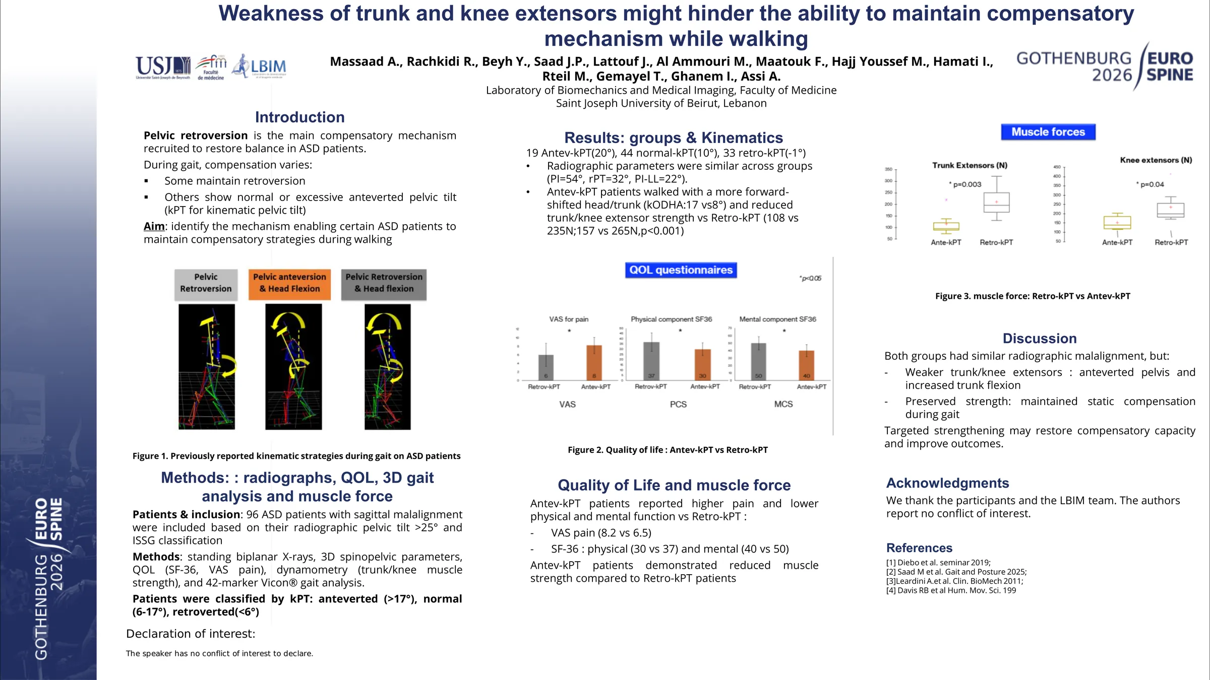
ASD patients are known to develop compensatory mechanisms in response to their sagittal malalignment. While pelvic retroversion is commonly recruited as a compensatory strategy in the standing position, functional evaluation in ASD has recently shown that some patients maintain pelvic retroversion during walking, while others adopt a normal or anteverted dynamic pelvic tilt (dynPT). Therefore, the aim of this study was to investigate the underlying mechanisms that enable certain patients to maintain compensatory mechanisms while walking.
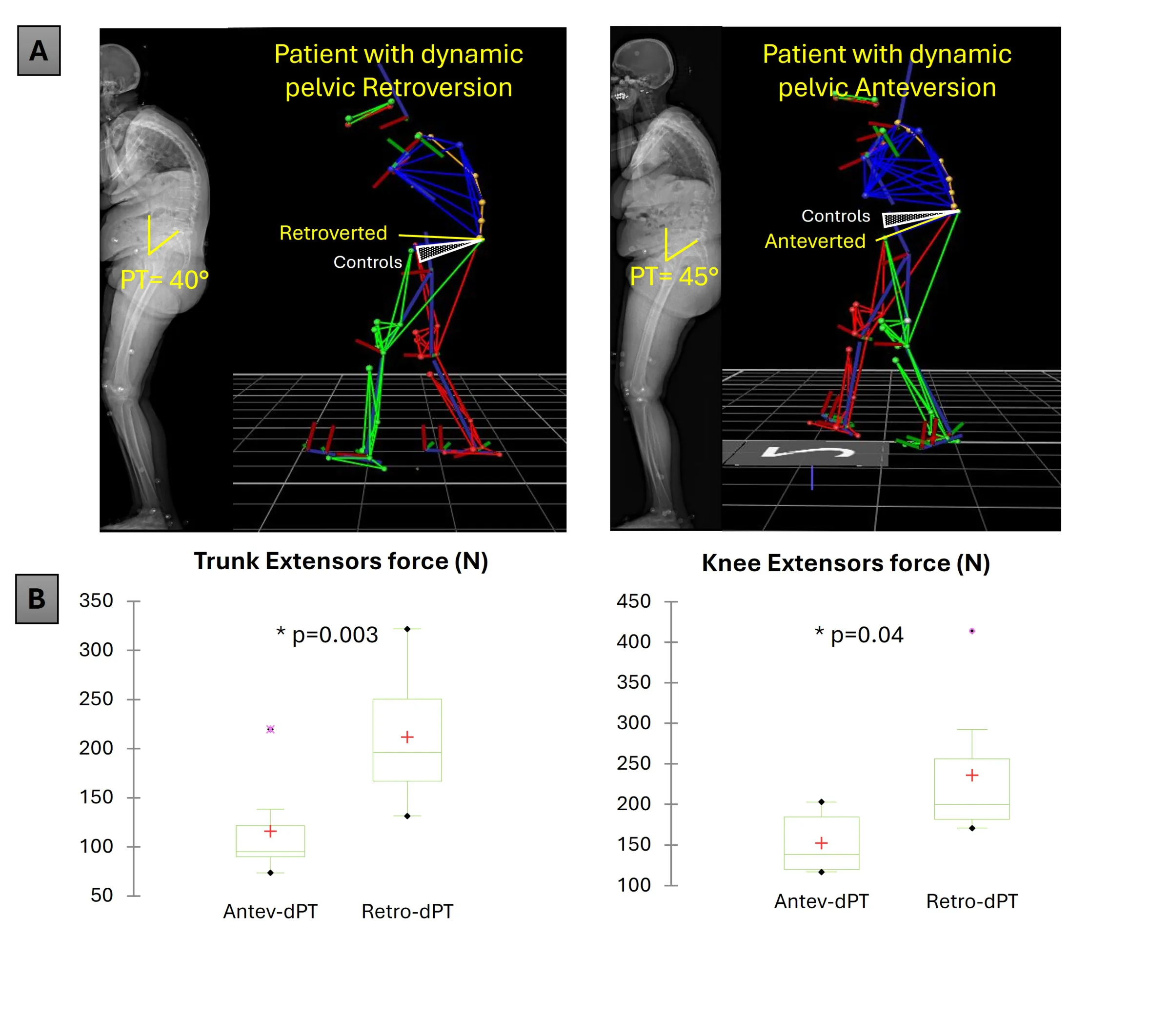
ASD patients underwent biplanar X-rays with the calculation of 3D spinopelvic parameters and filled QOL questionnaires (SF36, VAS for pain, Beck’s Depression Inventory BDI). Only patients with sagittal malalignment (SVA>50mm and/or PI-LL>10°) and having a radiographic PT>25° were included. Trunk and lower limb muscle forces were captured using a handheld dynamometer. They all underwent 3D gait analysis from which spine, pelvis and lower limb kinematics were calculated. Patients were classified depending on their dynPT behavior relatively to a normative database of 150 controls: anteverted (Antev-dynPT <-17°), normal (-17 to -6°) or retroverted (Retro-dynPT >-6°, Fig1.A). Comparisons were computed between groups.
96 ASD were included: 19 were classified as Antev-dynPT (-20°), 44 as normal-dynPT (-10°) and 33 as Retro-dynPT (1°). Radiographic spinopelvic parameters were similar between the 3 groups (PI:54°, PT:32°, SVA:75mm, PI-LL:22°). Patients in the Antev-dynPT group walked with a forward shifted trunk and head (dynSVA: 177 vs 122mm and dynODHA: 17 vs 8°) and exhibited a reduced trunk and knee extensor forces compared to the Retro-dynPT group (108 vs 235N, 157 vs 265N resp., all p<0.001; Fig1.B). Patients in the Antev-dynPT group reported more deteriorated QOL scores compared to the Retro-dynPT group (VAS: 8.2 vs 6.5, physical component SF36: 32 vs 37, BDI: 17 vs 10, all p<0.05).
Despite comparable radiographic sagittal malalignment, ASD patients with weak trunk and knee extensors tend to walk with an anteverted pelvis and increased trunk flexion. In contrast, patients with preserved muscle capacity appear to maintain their static compensatory mechanisms during gait. Personalized muscle strengthening programs may restore patients' ability to utilize compensatory mechanisms while walking, thereby enhancing surgical and non-surgical outcomes.
Figures and tables
As submitted with the abstract. Tap a figure to open it at full size.