
Growing Spine
Ultrasound Assessment of Static and Dynamic Spinal Flexibility in Mild to Moderate Adolescent Idiopathic Scoliosis
- The Hong Kong Polytechnic University, Hong Kong, China
- The University of Hong Kong (HKU), Hong Kong, China
- The Hong Kong Polytechnic University, Hong Kong, Hong Kong
Abstract
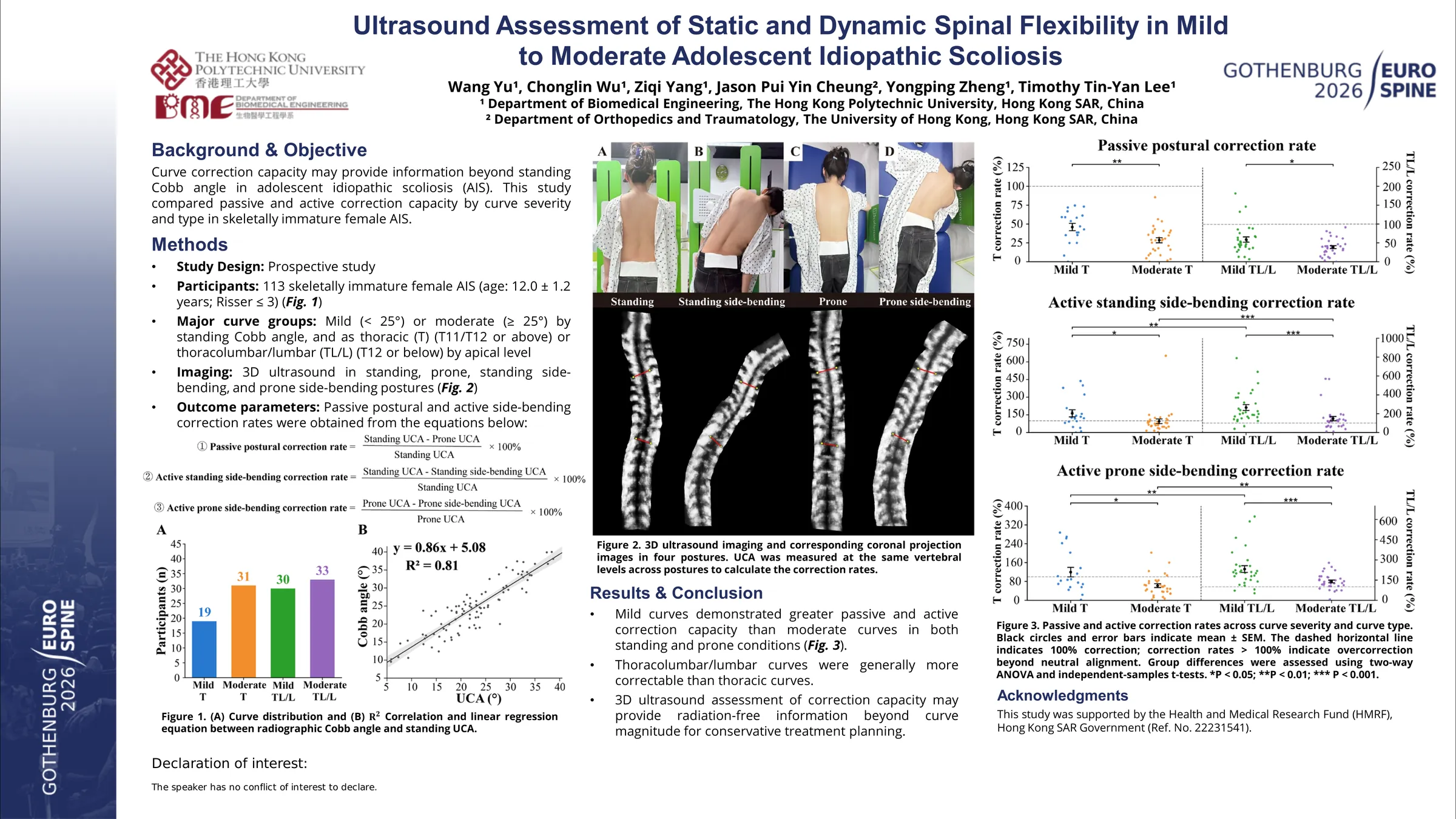
While clinical guidelines recommend bracing for adolescent idiopathic scoliosis (AIS) curves exceeding 20° and observation for those under 15°, recent evidence shows skeletally immature patients with small curves may progress at rates comparable to larger curves, suggesting that observation alone may be inadequate for mild cases. Spinal flexibility has been identified as a robust predictor of curve progression, highlighting the potential value of early assessment. Three-dimensional (3D) ultrasound offers a radiation-free alternative modality for multi-positional assessment of spinal deformity, therefore, this study aimed to evaluate the static and dynamic spinal flexibility in skeletally immature AIS patients by 3D ultrasound.
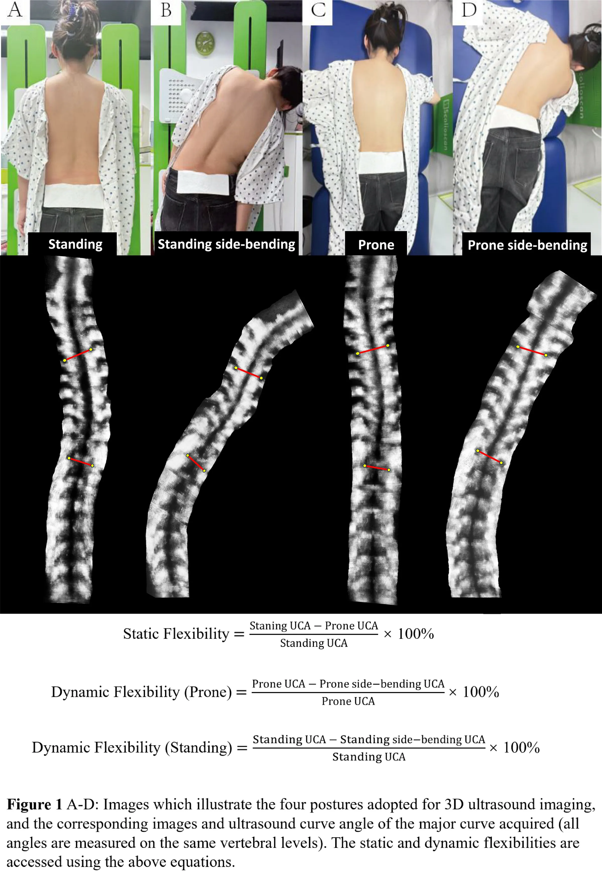
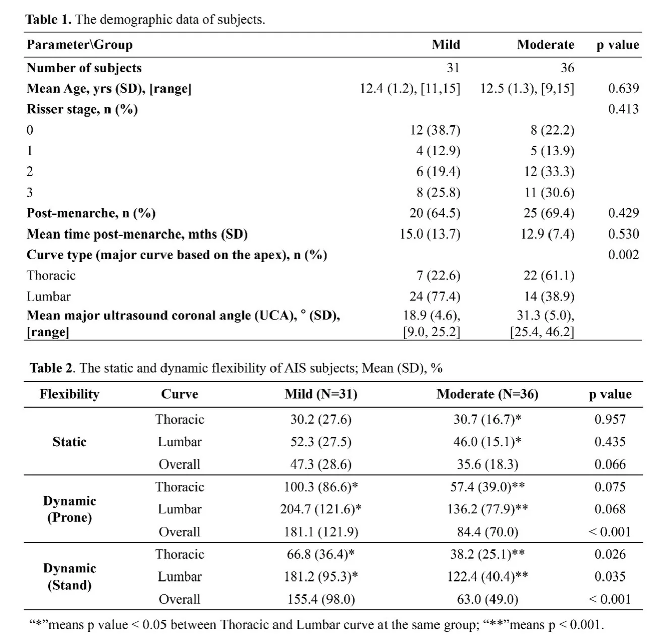
67 female AIS subjects (Risser sign ≤ 3) were prospectively recruited (Table 1). Ultrasound curve angle (UCA), closely correlated with the radiographic Cobb angle, was used to categorize the major curve severity into mild (< 25°, n = 31) and moderate (≥ 25°, n = 36) groups. Subjects underwent 3D ultrasound scanning in four positions: standing, prone, standing and prone side-bending to the convex side (Figure 1). Static flexibility represented correction from standing to prone, while dynamic flexibility reflected active correction toward the convex side during lateral bending. Group comparisons used t-tests and Chi-square tests, two-way ANOVA examined interactions between curve severity and type.
Overall, the mild group demonstrated greater static flexibility (47.3% vs. 35.6%, p = 0.066) and dynamic flexibility in both prone (181.1% vs. 84.4%, p < 0.001) and standing (155.4% vs. 63.0%, p < 0.001) positions compared to the moderate group (Table 2). The moderate group had a significantly higher proportion of thoracic curves than the mild group (p = 0.002). Subgroup analysis revealed that lumbar curves exhibited significantly greater static (p = 0.009) and dynamic (p < 0.001) flexibility than thoracic curves within moderate group. Similarly, mild group exhibited greater dynamic flexibility (p < 0.05) in the lumbar curves than in the thoracic curves.
Mild curves in skeletally immature AIS patients were demonstrated greater flexibility than moderate curves, with lumbar curves showing higher flexibility than thoracic ones, which may be attributable to the structural constraints of the rib cage. Nevertheless, dynamic flexibility provided a more distinct differentiation between mild and moderate curves compared to static assessment, suggesting its superiority for evaluating curve flexibility, but further validation through ongoing recruitment and longitudinal follow-up are warranted. Overall, ultrasound-based flexibility assessment enables region-specific profiling, which may enhance prognostic evaluation, inform conservative treatment planning, and reduce the need for additional radiographs in adolescent patients.
Figures and tables
As submitted with the abstract. Tap a figure to open it at full size.