
Cervical & Neural Pathologies
Trajectory Matters: Medial Angulation Improves Feasibility and Safety of C1 Pedicle Screw Placement
- BMC, Boston, United States of America
- Boston University School of Medicine, Hopkinsville, United States of America
- Boston University School of Medicine, Boston, United States of America
- Case Western Reserve University, Cleveland, United States of America
- University of Alabama at Birmingham, Birmingham, United States of America
- Cooper Medical School of Rowan University, Camden, United States of America
Abstract
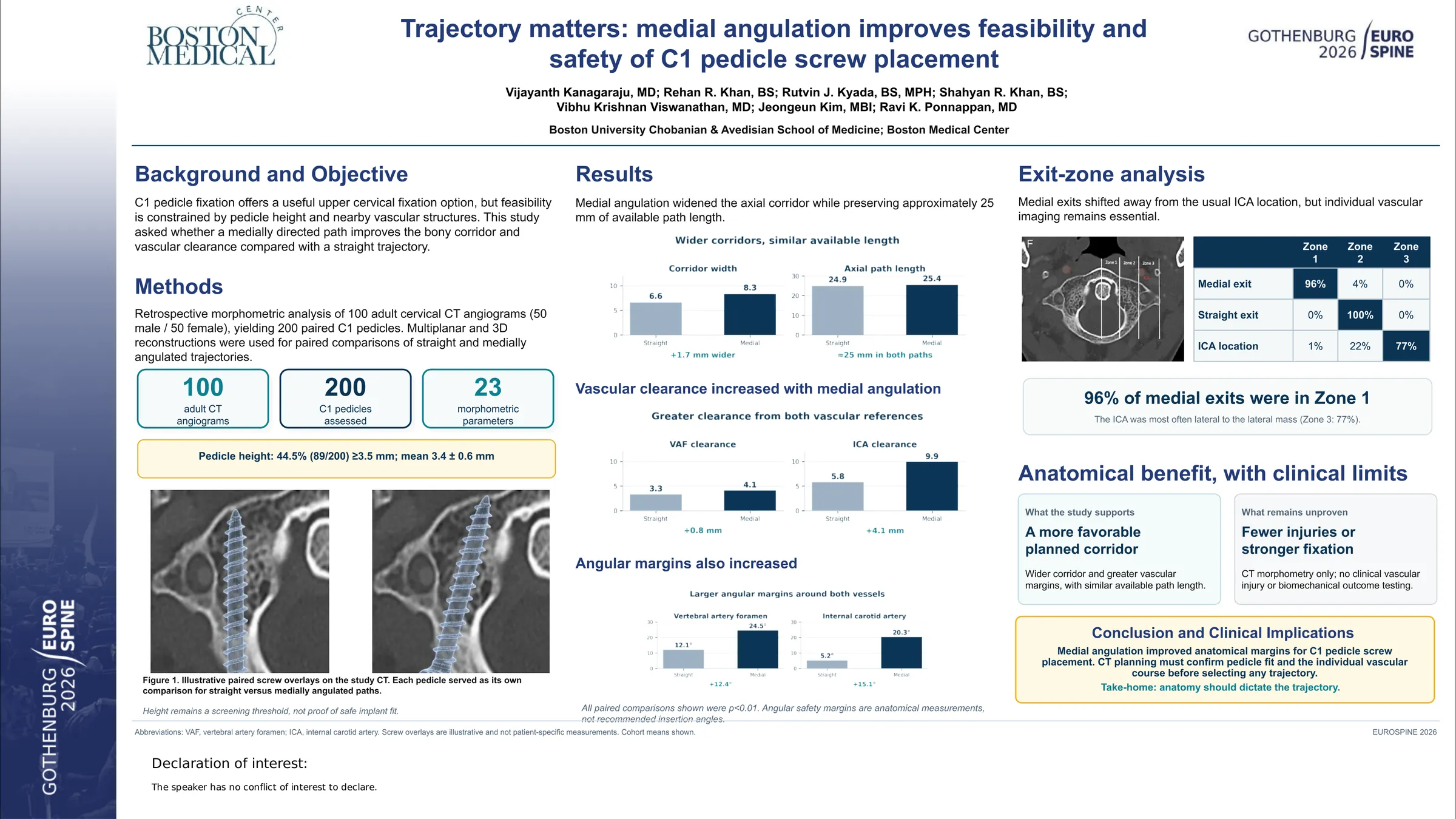
C1 pedicle screw fixation is able to provide robust atlantoaxial stability, although it carries a risk of neurovascular injury from narrow osseous corridors and proximity of the vertebral and internal carotid arteries. Additionally, population-tailored fixation feasibility remains of importance as the morphology of the atlas varies among different ethnic populations. South Asian populations have previously shown to have smaller on average C1 dimensions than Western populations. We sought to determine the feasibility of C1 pedicle screw fixation in an adult Indian cohort and also to compare the safety profiles of two C1 pedicle trajectories, straight (S) and medially angulated (M).
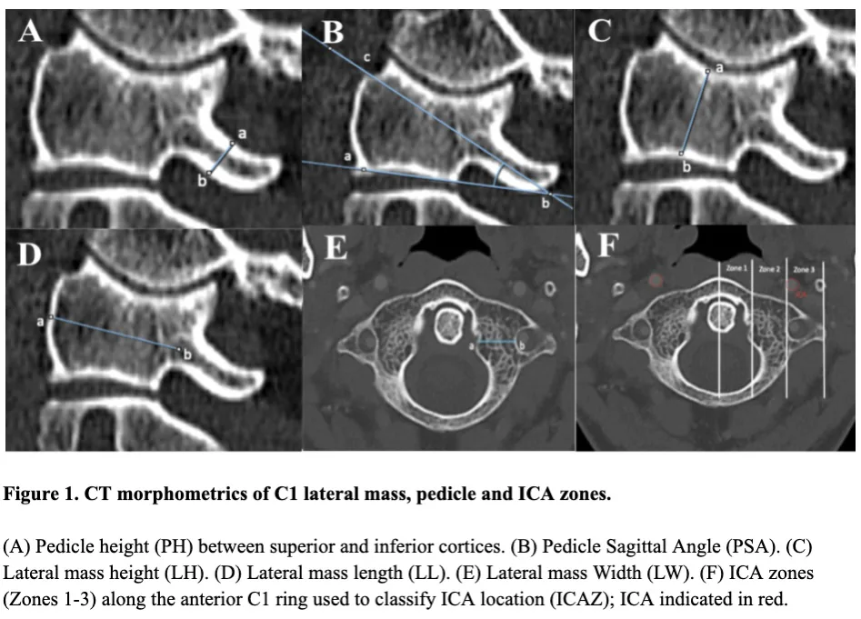
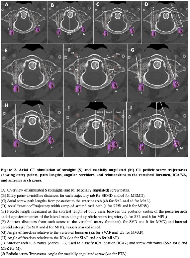
In this CT-based morphometric study, one hundred adult cervical CT angiograms (50 males, 50 females, 200 C1 pedicles) were reconstructed in multiplanar and three-dimensional formats for morphometric interpretation and analyses. A total of twenty-three morphometric parameters were measured for both S and M C1 pedicle screw trajectories. Outcomes measured comprised of C1 Pedicle morphometry/dimensions, safe screw corridor trajectory, path lengths, trajectory clearance from the vertebral artery foramen (VAF) and from the internal carotid artery (ICA), margin of angular safety, and the trajectory zone of exit. Direct comparisons were made between the S and M cohorts for each parameter, with sex-based subgroup analyses.
C1 pedicles were predominantly narrow and had an average height of 3.4 ± 0.6 mm. Only 44.5% were able to accommodate a standard 3.5-mm screw. M trajectories consistently produced wider screw corridors (8.3 mm vs 6.6 mm), greater clearance from the VAF (4.1 mm vs 3.3 mm) and ICA (9.9 mm vs 5.8 mm), and larger angular safety margins relative to both blood vessels (VAF: 24.5° vs 12.1°; ICA: 20.3° vs 5.2°; all p<0.01). Both trajectories had equivalent fixation path lengths with both preserving bicortical purchase. The ICA was most commonly found lateral to the lateral mass, 77%, while M screw trajectories exited predominantly within the safest medial zone, 96%, with its trajectory anchored away from the typical surrounding vasculature. Males demonstrated significantly larger osseous dimensions and safety corridors than females (p<0.01).
Standard C1 pedicle screw fixation is frequently constrained by narrow pedicle anatomy in adult Indian patients. M trajectories were found to optimize safe osseous corridors and neurovascular clearance for C1 fixation, without minimizing the fixation length. CT-based M trajectories may be safely performed in patients where C1 pedicle anatomy may be constrained.
Figures and tables
As submitted with the abstract. Tap a figure to open it at full size.