
Degenerative Thoracolumbar
The Quadratus Lumborum in Chronic Low Back Pain: Insights from Imaging, Function, and Clinical Measurements
- Charité – Universitätsmedizin Berlin, Berlin, Germany
- Julius Wolff Institute for Biomechanics and Musculoskeletal Regeneration, Berlin, Germany
Abstract
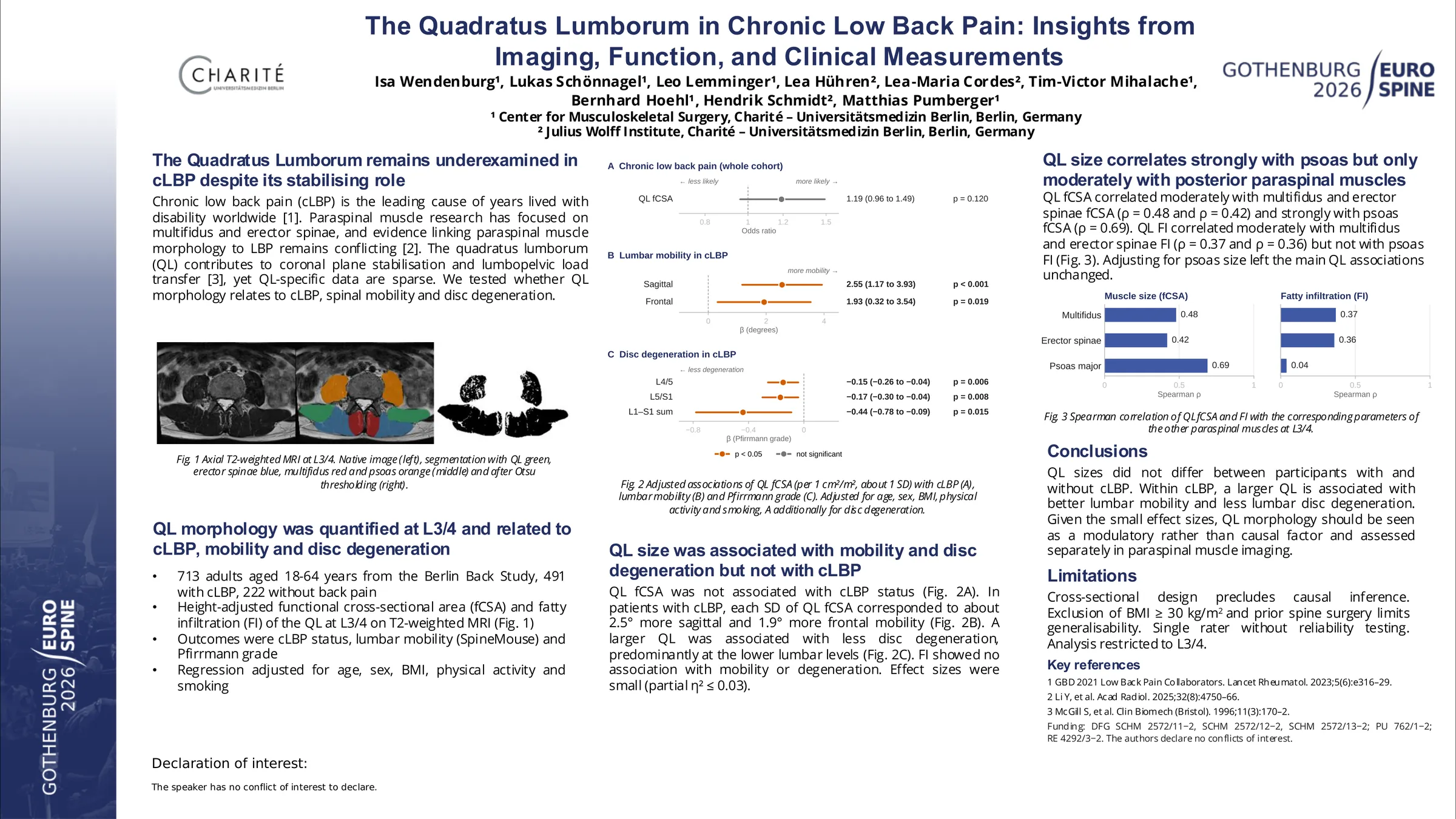
Chronic low back pain (cLBP) is the leading global cause of functional impairment and work disability. The quadratus lumborum muscle (QL) is considered an important stabilizer of the lateral and posterior lumbar spine. However, its structural relevance in cLBP remains insufficiently explored. This study aimed to assess morphological parameters of the QL using MRI and analyze their associations with pain, disability, spinal mobility, and lumbar degeneration.
Patients with and without cLBP were prospectively enrolled from the general population, excluding patients with fractures, tumors, infections or patients with previous spinal surgery. The QL was assessed in axial T2 weighted MRIs at all lumbar levels, with the level with the largest cross-section area (L3/4) selected for analysis. Height adjusted functional cross-sectional area (fCSA) and fatty infiltration (FI) of the QL were analyzed. To analyze the effect in the cLBP cohort, the association with pain intensity (von Korff Chronic pain grade), quality of life (SF-36), spinal mobility in the sagittal and frontal plane and lumbar disc degeneration (measured by the Pfirrmann Grade) were evaluated, adjusted for age, sex, BMI, physical activity and smoking status. Additionally, a sex specific analysis was conducted.
A total of 713 patients remained after exclusions, 222 (31.1%) without LBP and 491 (68.9%) with cLBP, with a mean age of 41.9 (SD 12.1) (Table 1). In the cLBP group, significant associations were found between QL fCSA and lumbar sagittal (β: 2.56, p < 0.001) and frontal (β: 1.92, p = 0.019) mobility as well as disc degeneration at the L4/5 and L5/S1 level (β: -0.15, p = 0.006 and β: -0.17, p = 0.009) and pain intensity (β: 5.21, p = 0.032) after accounting for confounders. Sex specific subgroup analysis demonstrated a stronger association of these findings in females, as well as an association with the physical component score of the SF-36 (β: 1.93, p = 0.021) (Figure 1). Effect sizes for the associations ranged from small to moderate (η² = 0.014 - 0.033).
QL morphology did not differ between individuals with and without cLBP and was not independently associated with pain status after adjustment for confounders. However, within symptomatic participants QL morphology showed consistent associations with spinal mobility and lumbar disc degeneration, with more significant effects in women. Given the small to moderate effect sizes, QL morphology should be interpreted as a modulatory rather than causal factor in cLBP. Imaging findings alone appear insufficient to characterise clinical status, and functional assessment may provide additional value. Future studies are needed to clarify the relationship between QL morphology, functional impairment, and degenerative changes, and to determine whether targeted conditioning of the QL contributes to improved outcomes in selected patient groups.
Figures and tables
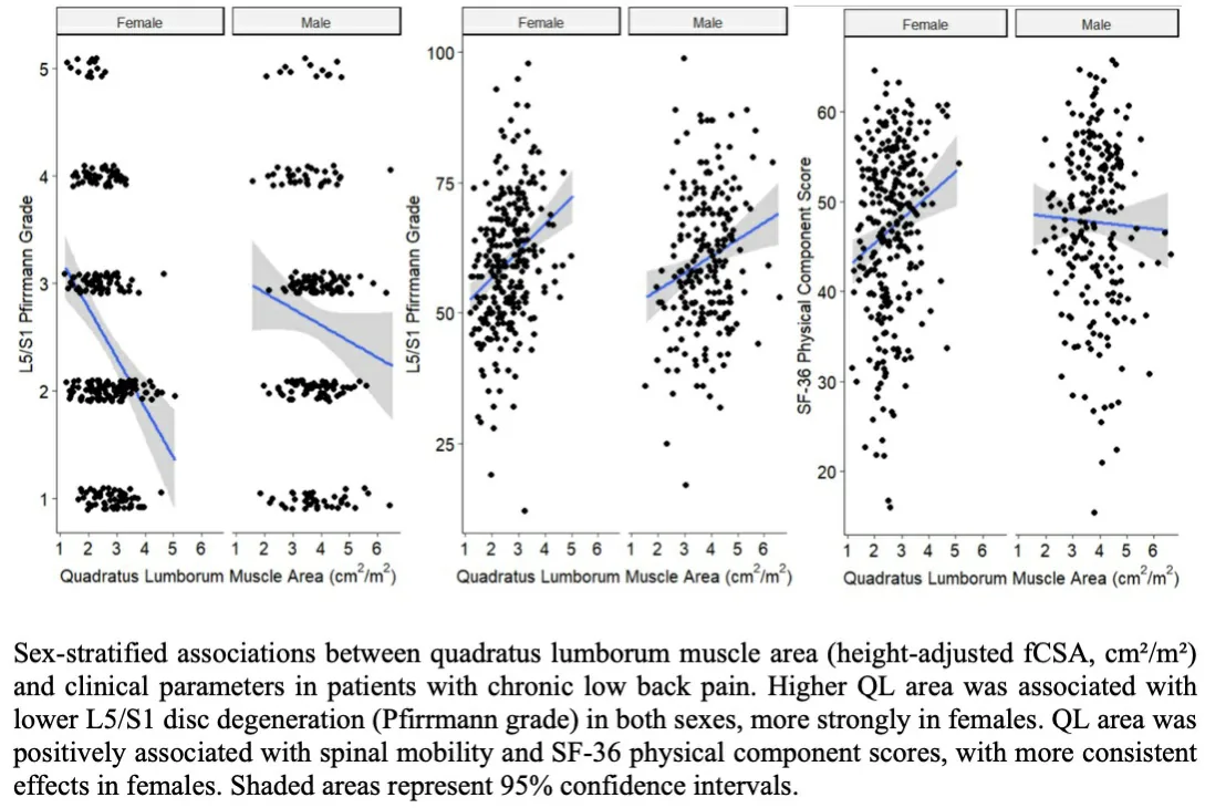
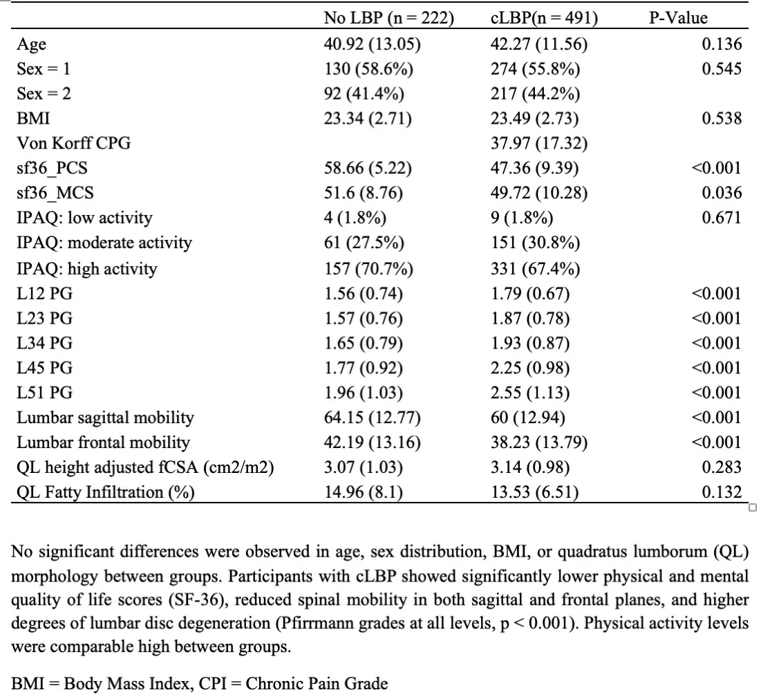
As submitted with the abstract. Tap a figure to open it at full size.