
Degenerative Thoracolumbar
The Potential of 3D MRI for Improved Detection and Evaluation of the Conjoined Lumbosacral Nerve Root
- Hospital for Special Surgery, New York, United States of America
- Hospital for Special Surgery Main Hospital, New York, United States of America
- Hospital for Special Surgery Main Hospital, Zürich, United States of America
- Universitätsklinik Balgrist, zurich, Switzerland
Abstract
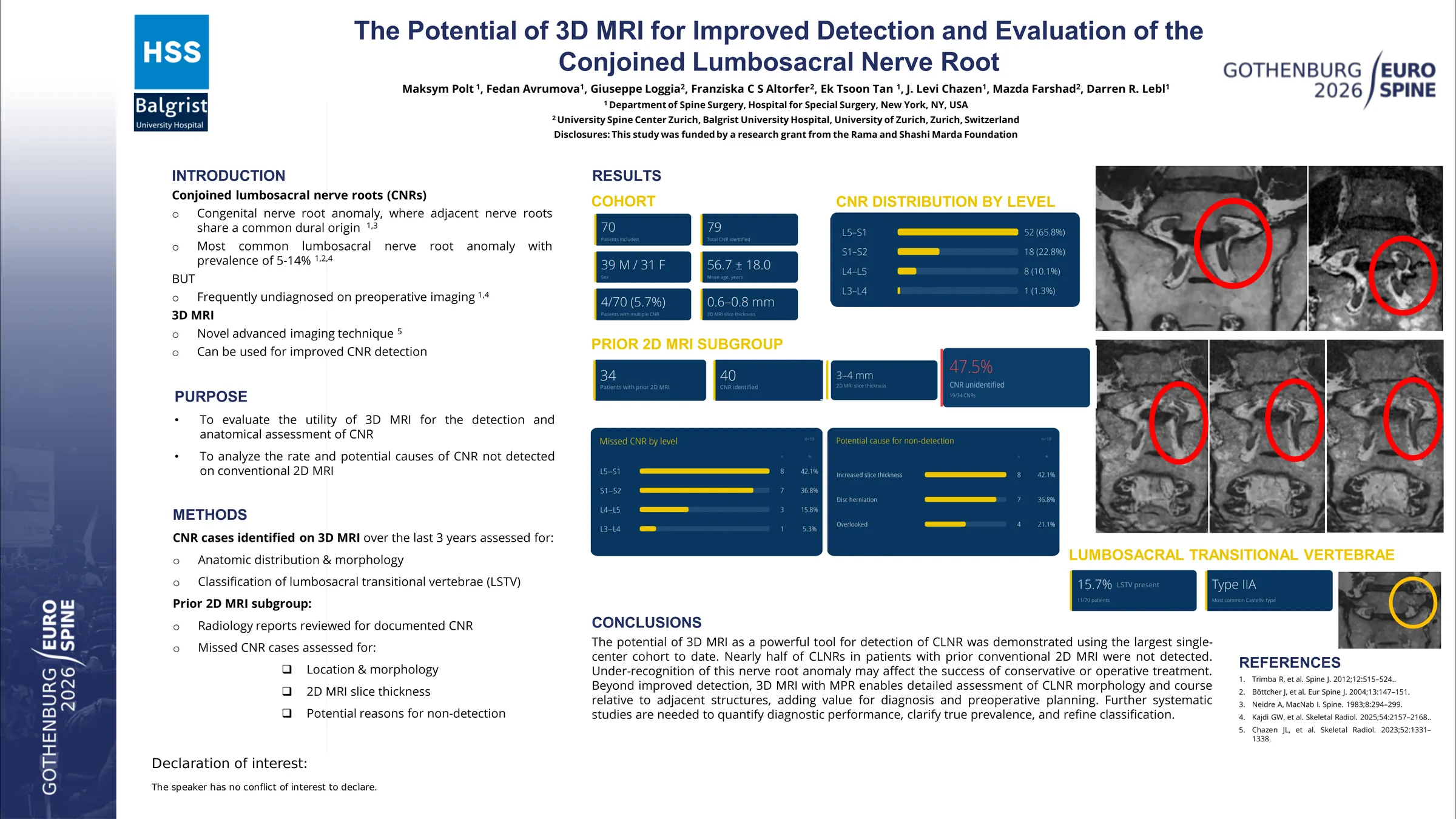
Conjoined lumbosacral nerve roots (CLNR) are often missed during diagnostic work-up, increasing the risk of inappropriate treatment, iatrogenic injury, or failed back surgery. Conventional two-dimensional (2D) magnetic resonance imaging (MRI) tends to demonstrate modest diagnostic accuracy, as slice thicknesses of 3-4 mm and reliance on separate 2D planes may limit assessment of the CLNR course. Three-dimensional (3D) MRI provides a single volumetric dataset with sub-millimeter slice thickness, potentially improving evaluation of subtle structures such as CLNR. This study evaluates the utility of 3D MRI in the detection and anatomical assessment of CLNR, while analyzing the rate and potential causes of CLNR not detected on conventional 2D MRI.
A retrospective search of the institutional imaging database was performed to identify patients with CLNR detected on 3D MRI over the past 3 years. Patient demographics and anatomic distribution were recorded. Furthermore, CLNR was classified according to Neidre and McNab, and the presence of lumbosacral transitional vertebrae (LSTV) was assessed using the Castellvi classification. Multiplanar reconstruction (MPR) was used for detailed anatomical assessment. A subgroup with prior conventional 2D MRI was identified, and corresponding radiology reports were reviewed to determine whether CLNR had been reported. Cases in which CLNR was not detected on 2D MRI were further analyzed for anatomic localization, slice thickness, and possible reasons for undetected CLNR on 2D MRI.
Within 70 patients (mean age 56.7 ± 18.0, 39 males and 31 females) 79 CLNRs were identified using a 3D MRI (slice thickness of 0.6 - 0.8 mm). CLNR was most commonly found at the L5-S1 level (65.8%), followed by S1-S2 (22.8%), L4-L5 (10.1%), and L3-L4 (1.3%). According to the Neidre and McNab classification, 23 (29.1%) cases fell into type IA and 50 (63.3%) into type IB, with 6 (7.6%) variants not covered by the classification. LSTV was found in 11 (15.7%) patients, with Castellvi type IIa being most prevalent. Among 70 patients, 34 (40 CLNR) had undergone 2D MRI prior to 3D MRI. With 2D MRI slice thicknesses of 3-4 mm, 19 (47.5%) CLNR went undetected. Of these, 1 (5.3%) nerve root anomaly occurred at L3-L4, 3 (15.8%) at L4-L5, 8 (42.1%) at L5-S1, and 7 (36.8%) at S1-S2. The most common potential causes of undetected CLNR on 2D MRI included increased slice thickness and disc herniation obscuring or compressing the nerve roots.
The potential of 3D MRI as a powerful tool for detection of CLNR was demonstrated using the largest single-center cohort to date. Nearly half of CLNRs were not detected on prior 2D MRI, which may compromise conservative or surgical treatment. Beyond improved detection, 3D MRI with MPR enables detailed assessment of CLNR morphology and course relative to adjacent structures, adding value for diagnosis and preoperative planning.
Figures and tables
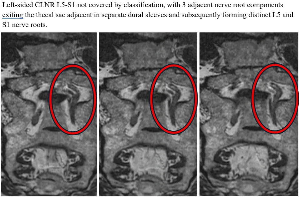
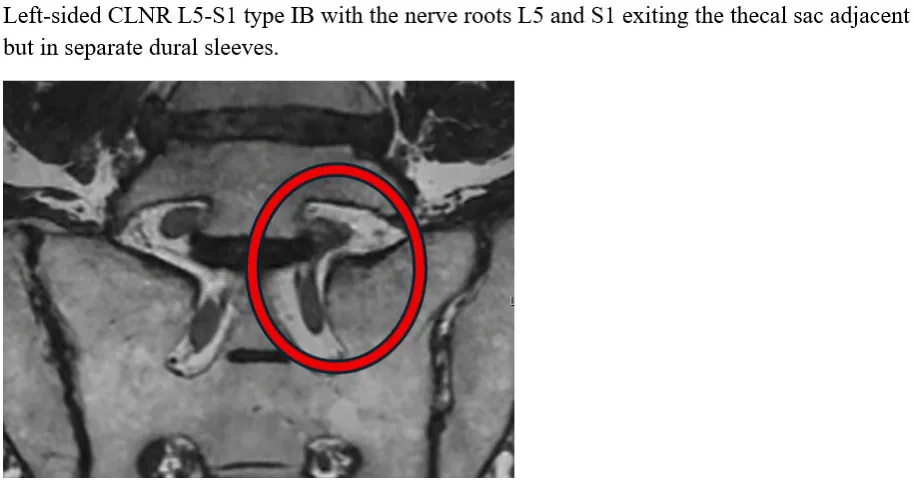
As submitted with the abstract. Tap a figure to open it at full size.