
Cervical & Neural Pathologies
Comparison of Osteolysis and Revision Rates Following Anterior Cervical Discectomy and Fusion: Stand-Alone Cage Versus Cage-Screw Construct
- Norfolk and Norwich University Hospital, Oswestry, United Kingdom
- Norfolk and Norwich University Hospital, Norwich, United Kingdom
Abstract
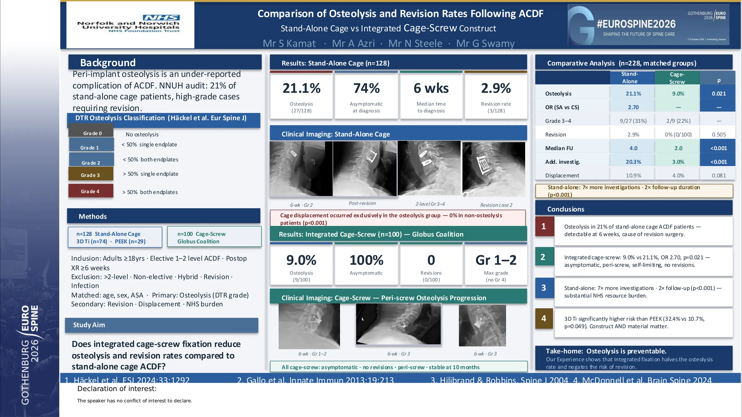
It was noticed by our department significant cases of revision post standalone cages in ACDF surgery secondary to septic osteolysis. On further investigation rates of aseptic osteolysis were shown to be higher and also the need for additional investiogations and longer follow ups increasing the economic burden on the hospital and shift was made to a cage screw construct. This study is to compare the rates of aseptic osteolysis and subsequent revision rate in ACDF surgery.
A retrospective comparative cohort study was performed at Norfolk and Norwich Hospital from 2019-2025. Patients undergoing primary one- to three-level ACDF with either a stand-alone cage or a cage-screw construct were included if postoperative imaging was available with minimum follow up of 6 weeks. Osteolysis was defined as focal progressive radiolucency adjacent to the implant and graded according to severity. Revision surgery, subsidence, and requirement for additional postoperative investigations and follow up period were recorded. Outcomes were compared descriptively between constructs.
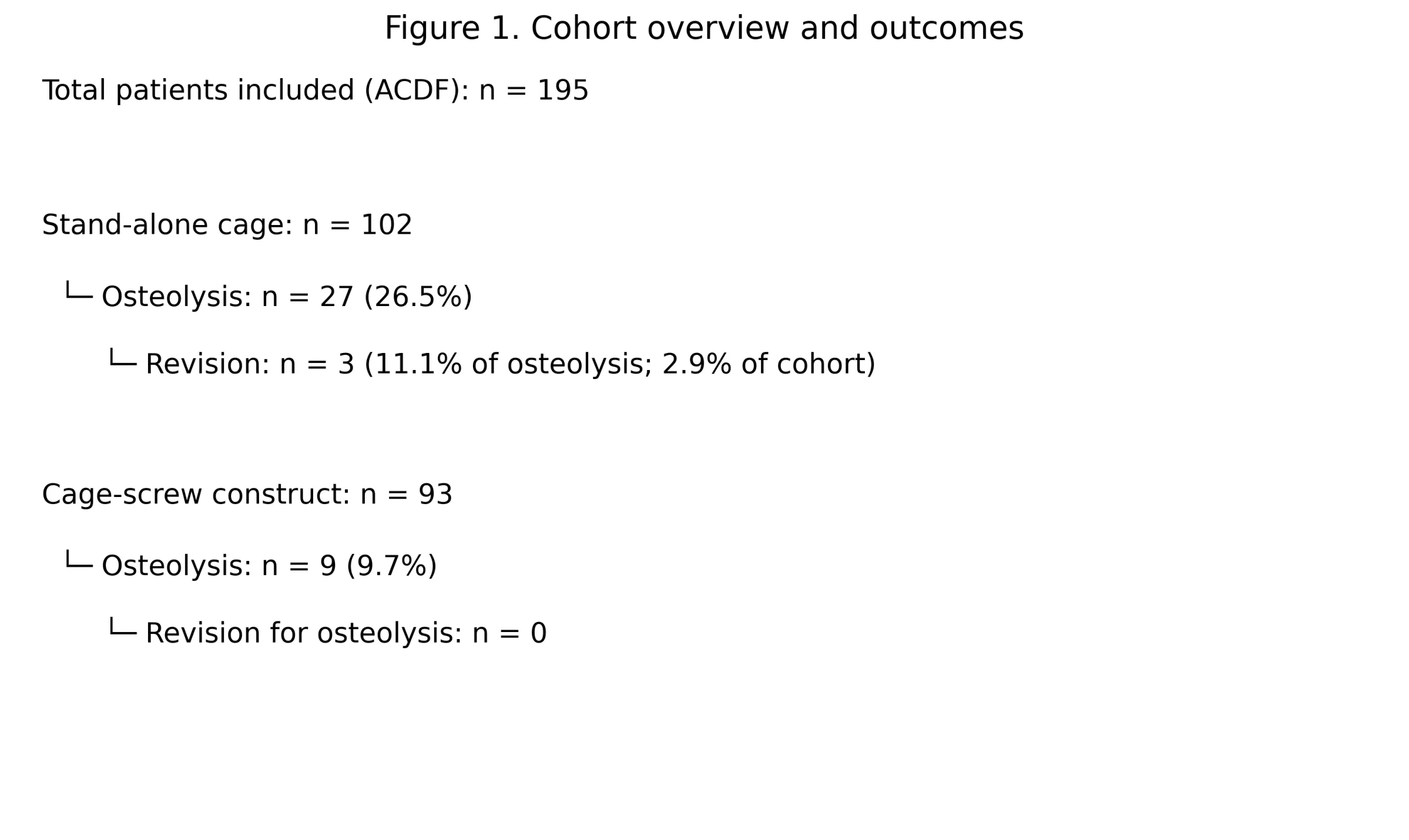
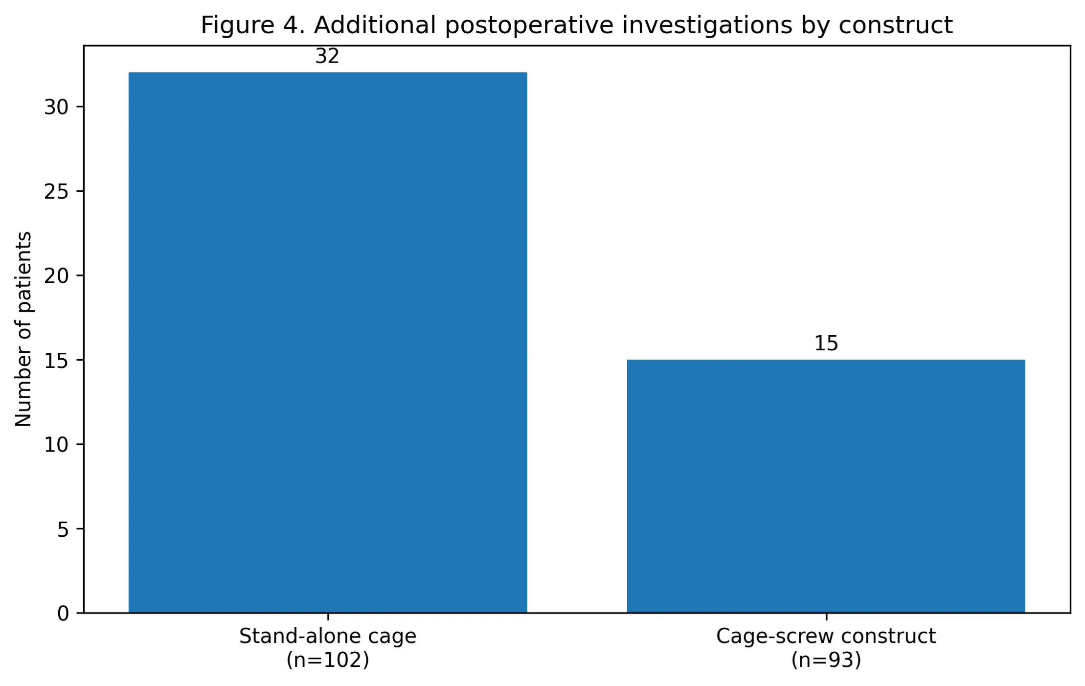
A total of 195 patients were included (102 stand-alone cage, 93 cage-screw construct). Mean age was comparable between patients with and without osteolysis (66.3 vs 65.2 years). The mean number of operated levels was similar in both groups (1.26 vs 1.27) Osteolysis was more frequently observed in the stand-alone cage group (~21%) compared with the cage-screw construct group (9.7%). Osteolysis following stand-alone cages demonstrated a wider range of severity, including higher-grade cavitary changes, whereas osteolysis in the cage-screw construct group was predominantly Grade 1, with only one case of Grade 3 osteolysis associated with mild segmental angulation. The overall revision rate was higher in the stand-alone cage group (2.9%), with all revisions occurring in patients with osteolysis, while no revisions for osteolysis occurred in the cage-screw construct group. Patients in both groups with osteolysis required increased postoperative surveillance, with additional cross-sectional imaging performed in 15 patients in the cage-screw construct group. Mean follow-up was longer in the stand-alone cage cohort compared with the cage-screw construct cohort
Osteolysis occurs more frequently and with greater severity following ACDF using stand-alone cages compared with cage-screw constructs and is associated with a higher revision rate confined to the stand-alone cohort. It was also noted that need of additional investigations and follow ups were higher in stand alone cages as compared to cage-screw construct making a cage screw construct economical better as compared to stand alone cages.These findings suggest implant construct may influence both the incidence and clinical consequences of osteolysis following ACDF.
Figures and tables
As submitted with the abstract. Tap a figure to open it at full size.