
Degenerative Thoracolumbar
Reperfusion blood oxygen level dependent (BOLD) magnetic resonance imaging (MRI) as a non invasive tool to distinguish neurogenic and vascular claudication
- Institution of Clinical Sciences, Sahlgrenska Academy, University of Gothenburg, Sweden, Gothenburg, Sweden
- Institute of Clinical Sciences, Sahlgrenska Academy, University of Gothenburg, Gothenburg, Sweden
- Institute of Medicine, Sahlgrenska Academy, University of Gothenburg, Gothenburg, Sweden
Abstract
Patients with lumbar spinal stenosis (LSS) and patients with peripheral arterial occlusive disease (PAOD) both experience claudication symptoms (neurogenic or vascular claudication). These conditions may occur simultaneously in different grades and it can sometimes be difficult to determine which is the primary diagnosis for the leg symptoms. BOLD-MRI relies on magnetic differences of oxygenated and deoxygenated haemoglobin, and regional changes of oxy/deoxyhaemoglobin ratio are recorded by dynamic T2* measurements. Non-invasive reperfusion BOLD-MRI of lower limb tissue have been shown to clearly distinguish healthy controls from patients with PAOD. The main aim here was to determine if lower limb reperfusion BOLD-MRI protocol could be used to differentiate LSS from PAOD patients.
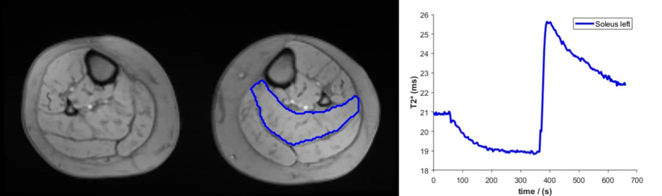
Eighteen patients with LSS (mean age 72.8 ± 6.8 SD years), without any clinical suspicion of PAOD, underwent lower limb BOLD-MRI and were compared with a previously reported cohort of PAOD patients (n=22, age73.5±4.0 y) and a healthy control cohort (n=10, age 67.9±6.2 y), examined at the same centre. All LSS patients underwent a repeated reperfusion BOLD-MRI following provoking of claudication by treadmill walking (same day) and assessments compared. For all groups a transverse slice of the calf was imaged repeatedly during an upper-thigh cuff compression cycle consisting of baseline (1-min), cuff inflated arterial occlusion (5-min) and cuff deflated reactive hyperaemia (5-min), Fig 1. Baseline, peak T2* and time-to-peak (TTP) after cuff deflation were assessed in the soleus muscle. Non-parametric Kruskal Wallis tests were used to test differences between patient groups/controls and Wilcoxon signed rank test for comparisons of LSS group repeated measurements.
There were no differences between groups for baseline T2*, the peak T2* nor for Δ peak T2* (baseline to peak T2*). Δ peak T2* percentage for the three groups were 13%, 9% and 8% for LSS, PAOD and controls respectively, and for walking provoked LSS 11%.
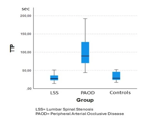
For the TTP there was a difference between LSS and PAOD, mean (SD) 29(10) and 100(42) (p<0.001), but no difference between LSS and controls 40(31) (p=0.33). Neither were there any difference between the two LSS group measurements, p=0.74), Fig. 2.
The LSS cohort differed from the PAOD cohort in TTP in a manner consistent with previous comparisons between healthy controls and PAOD patients. No individuals with both suspected LSS and PAOD were however included in the present study. Reperfusion BOLD MRI warrants further investigation in mixed or uncertain claudication etiology. Based on the present findings reperfusion BOLD MRI could represent a valuable non-invasive clinical tool for patients with unclear cause of claudication patients.
Figures and tables
As submitted with the abstract. Tap a figure to open it at full size.