
Complications & Epidemiology
Reoperation Rates and Radiographic Outcomes After 1- and 2-Level Standalone Lateral Lumbar Interbody Fusion for Adult Spinal Deformity
- Hospital for Special Surgery Main Hospital, New York, United States of America
Abstract
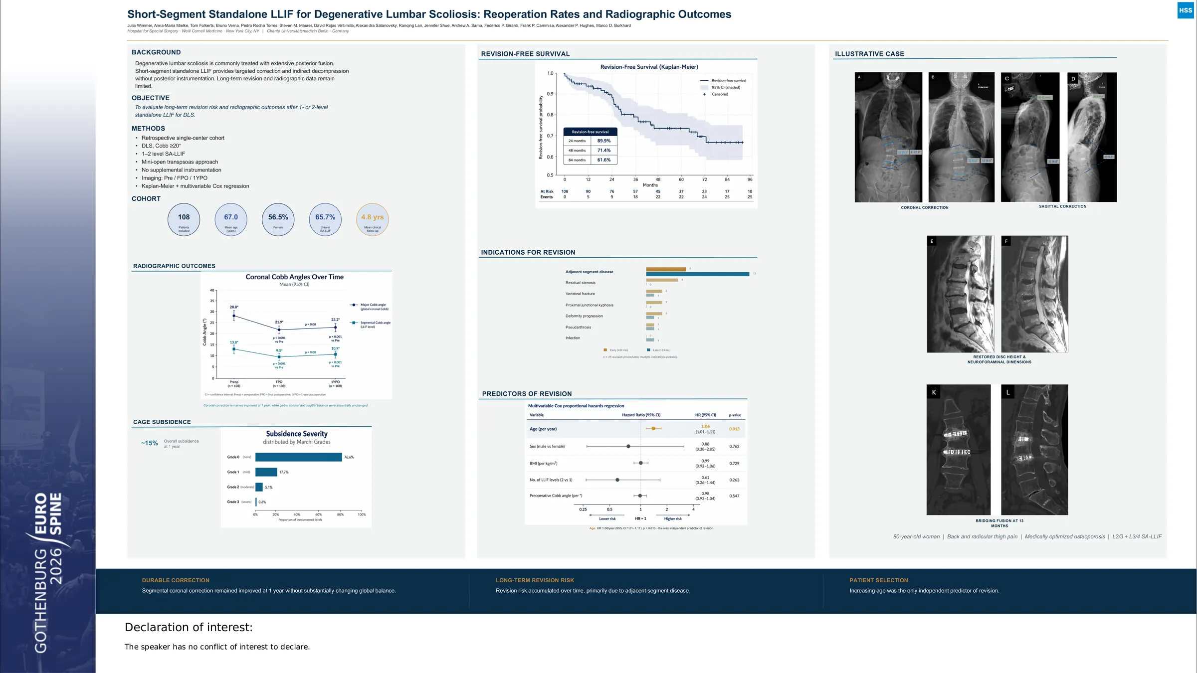
Degenerative lumbar scoliosis (DLS) often requires surgical treatment when conservative measures fail. Standalone lateral lumbar interbody fusion (SA-LLIF) has been increasingly used for short-segment correction in selected patients with DLS; however, evidence regarding durability and radiographic outcomes remains limited.
Patients with DLS and a preoperative lumbar Cobb angle ≥ 20° who underwent 1- or 2-level SA-LLIF at an academic spine center were retrospectively reviewed. A total of 108 patients were included, with a mean follow-up of 4.0 ± 3.3 years; 39 patients had a follow-up of less than 2 years. Radiographic parameters, including Cobb angle, coronal vertical axis (CVA), lumbar lordosis (LL), and sagittal vertical axis (SVA), were assessed preoperatively, at the first postoperative (FPO) radiograph, and at one year postoperatively (1YPO). Revision-free survival was analyzed using Kaplan-Meier methods, and predictors of revision were evaluated using multivariable Cox regression.
During follow-up, 23 patients (21.3%) underwent revision surgery. Revision-free survival was 89.9% at 24 months. Among the 10 revision procedures performed within the first 2 years, the most common indications were adjacent segment disease (n=6, 60%) and residual stenosis (n=4, 40%); multiple indications were present in some cases. Increasing age was the only variable independently associated with revision surgery (HR 1.07 per year, 95% CI 1.02-1.13; p = 0.005). Radiographically, Cobb angle improved from 28.8° preoperatively to 21.9° postoperatively and was maintained at 1YPO. LL increased from 42.4° preoperatively to 45.5° postoperatively and remained stable at 1YPO, while coronal vertical axis (CVA) and sagittal vertical axis (SVA) remained largely unchanged.
In patients with DLS undergoing 1- or 2-level SA-LLIF, the rate of revision surgery within the first 2 years was low, with cumulative revision rates over longer follow-up comparable to those reported for posterior fusion procedures. Revision risk was primarily influenced by patient age rather than radiographic parameters. Overall, these findings support SA-LLIF as a less invasive surgical option for carefully selected patients with DLS, achieving sustained coronal and sagittal correction with an acceptable reoperation profile.