EUROSPINE 2026 — Spine in Motion Gothenburg, 7–9 October 2026

Adult Deformity

Relationship Between Preoperative Cervical Muscle Morphology and Reciprocal Cervical Alignment Changes After Lumbar PSO

B. Verna1, M. Burkhard1, A. Guven1, A.M. Mielke1, J. Wimmer1, T. Folkerts1, P.A. Rocha Torres1, J. Zhu1, J. Shue1, A. Sama1, F. Girardi1, F. Cammisa1, A. Hughes1

  1. Hospital for Special Surgery Main Hospital, New York, United States of America
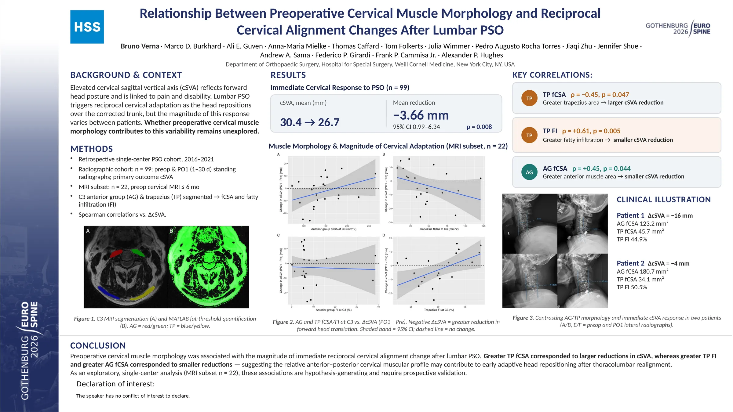
Poster 000499: Relationship Between Preoperative Cervical Muscle Morphology and Reciprocal Cervical Alignment Changes After Lumbar PSO
Abstract no.
000499
Topic
Adult Deformity
Session
ePoster - Adult Spinal Deformity
Author
B. Verna
Open full e-poster

Opens at full size in a new tab — zoom in to read the detail.

Abstract PDF

Abstract

Lumbar sagittal malalignment induces compensatory changes throughout the thoracic and cervical spine. Although lumbar correction procedures such as PSO are known to produce reciprocal changes in cervical alignment, the clinical determinants of this response remain poorly understood. Given the role of cervical musculature in stabilizing head posture and motion, cervical muscle morphology may influence these reciprocal alignment changes. The objective of our study is to evaluate whether preoperative cervical muscle morphology is associated with immediate cervical alignment changes following lumbar pedicle subtraction osteotomy (PSO).

Patients who underwent lumbar PSO with preoperative and immediate postoperative (1-30 days) standing whole-spine lateral radiographs were reviewed. C2-7 sagittal vertical axis (cSVA) was measured at both timepoints. For muscle analysis, a subset with an available preoperative cervical MRI was included. At C3, anterior muscle group (AG) and trapezius (TP) functional cross-sectional area (fCSA) and fatty infiltration (FI) were obtained.

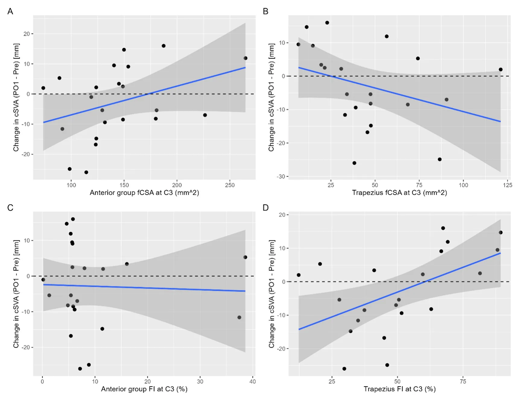
Ninety-nine patients were included in the initial analysis. Mean cSVA significantly decreased from 30.4 to 26.7 mm. Twenty-two patients had a preoperative cervical MRI available for muscle analysis. Spearman analysis demonstrated that greater TP fCSA correlated with larger postoperative reductions in cSVA (rho = -0.45, p = 0.047), whereas greater TP FI correlated with smaller reductions in cSVA (rho = 0.61, p = 0.005). Larger AG fCSA also correlated with smaller reductions in cSVA (rho = 0.45, p = 0.044).

Preoperative cervical muscle morphology was associated with reciprocal cervical alignment changes after PSO. Greater TP fCSA correlated with larger postoperative reductions in cSVA, whereas greater TP FI correlated with smaller reductions. Larger AG fCSA also correlated with smaller immediate cervical alignment changes. Together, these findings suggest that the relative balance between posterior stabilizing and anterior cervical muscle quality may influence the magnitude of acute cervical decompensation following thoracolumbar realignment.

Figures and tables
Representative examples of patients with contrasting cervical muscle morphologies and corresponding differences in immediate postoperative change in cervical sa
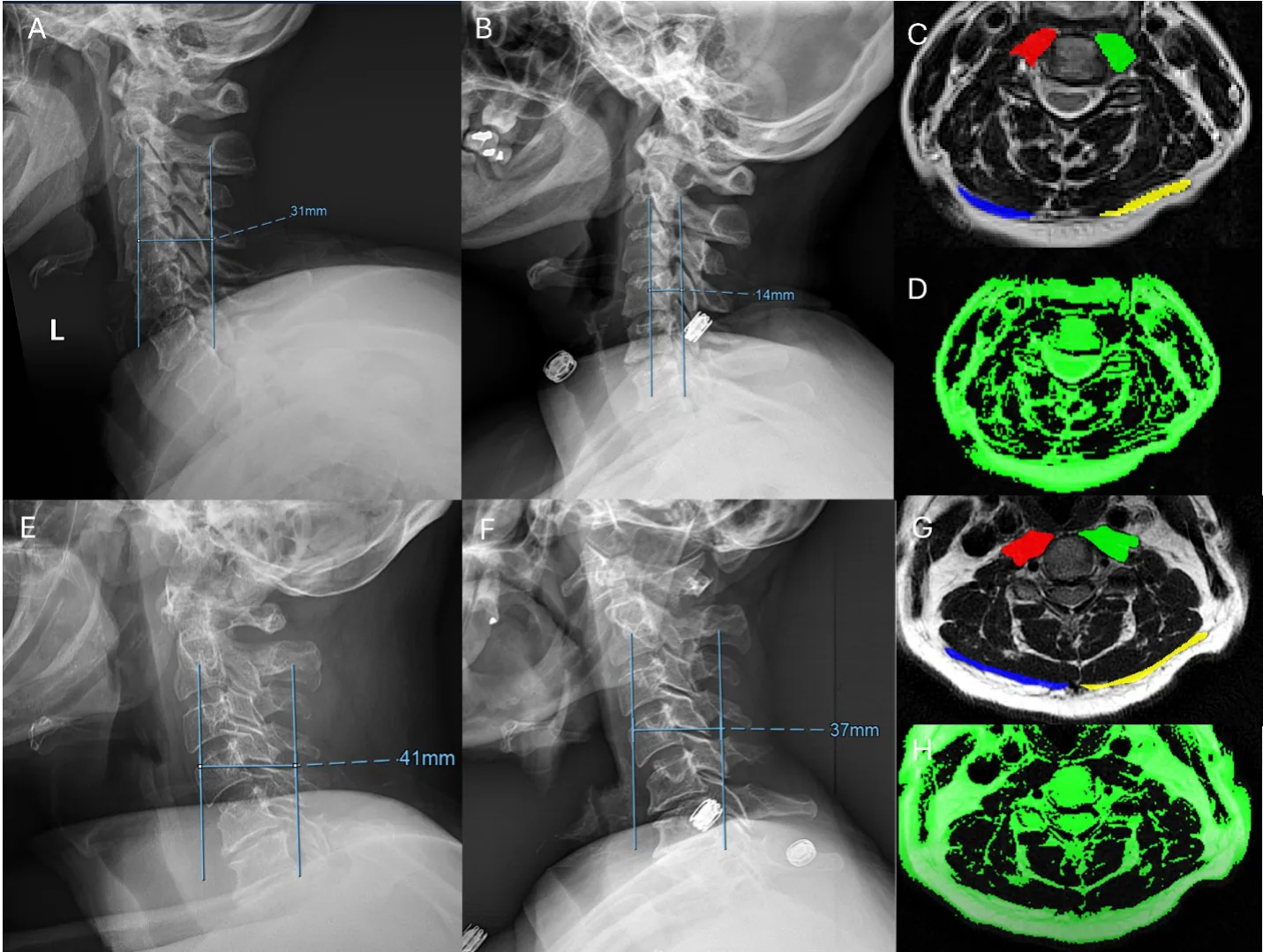
Representative examples of patients with contrasting cervical muscle morphologies and corresponding differences in immediate postoperative change in cervical sagittal vertical axis (ΔcSVA). Panels A-D show a 59-year-old male (Patient 1) with a larger decrease in cSVA from preoperative to immediate postoperative assessment (Pre: 31.0 mm; PO1: 14 mm; ΔcSVA = -16 mm), characterized by lower anterior group functional cross-sectional area (AG fCSA = 123.2 mm²), higher trapezius functional cross-sectional area (TP fCSA = 45.7 mm²), and lower trapezius fatty infiltration (TP FI = 44.9%). Panels E-H show a 57-year-old male (Patient 2) with a lower postoperative reduction in cSVA (Pre: 41 mm; PO1: 37 mm; ΔcSVA = -4 mm), characterized by higher AG fCSA (180.7 mm²), lower TP fCSA (34.1 mm²), and greater TP FI (50.5%). Notably, AG FI was similar between patients (AG FI: 5.41% vs 5.42%), consistent with the lack of association
observed between AG FI and ΔcSVA in the overall cohort. Preoperative and immediate postoperative alignment is shown using upright, standing whole-spine lateral
observed between AG FI and ΔcSVA in the overall cohort. Preoperative and immediate postoperative alignment is shown using upright, standing whole-spine lateral radiographs, and muscle composition is illustrated using segmentation-based visualization at the C3 level. Relationship between cervical paraspinal muscle morphology and immediate postoperative changes in cervical sagittal vertical axis (cSVA). Scatterplots illustrate the associations between anterior muscle group (AG) and trapezius (TP) functional cross-sectional area (fCSA) and fatty infiltration (FI) measured at C3, and the change in cSVA from preoperative to immediate postoperative assessment 1 (Change in cSVA = PO1 - Pre). Panels depict (A) AG fCSA, (B) TP fCSA, (C) AG FI, and (D) TP FI in relation to change in cSVA. Solid lines represent linear regression fits with 95% confidence intervals, and dashed horizontal lines indicate no change in cSVA (change in cSVA = 0).

As submitted with the abstract. Tap a figure to open it at full size.