
Adult Deformity
Reliability and Accuracy of Indirect T1 Slope Estimation Methods: A Three-Observer Radiographic Study of Cervical Sagittal Alignment
- San Juan de Reus University Hospital, Reus, Spain
Abstract
T1 slope is a key parameter of cervical sagittal balance; however, its direct measurement on lateral radiographs is not always feasible. Several indirect estimation formulas have therefore been proposed. The aim of this study was to compare the agreement and accuracy of the main available formulas.
Eighty-two patients who underwent surgery for cervical pathology between 2017 and 2025 were retrospectively analyzed. Preoperative standing lateral radiographs were reviewed to assess T1 visibility, cervical lordosis, and C2–C7 SVA. T1 slope was measured directly when possible and indirectly using established formulas based on C7 slope and cervical lordosis. MRI measurements performed within three months were also included. Three observers performed the measurements, and intra- and interobserver reliability were assessed.
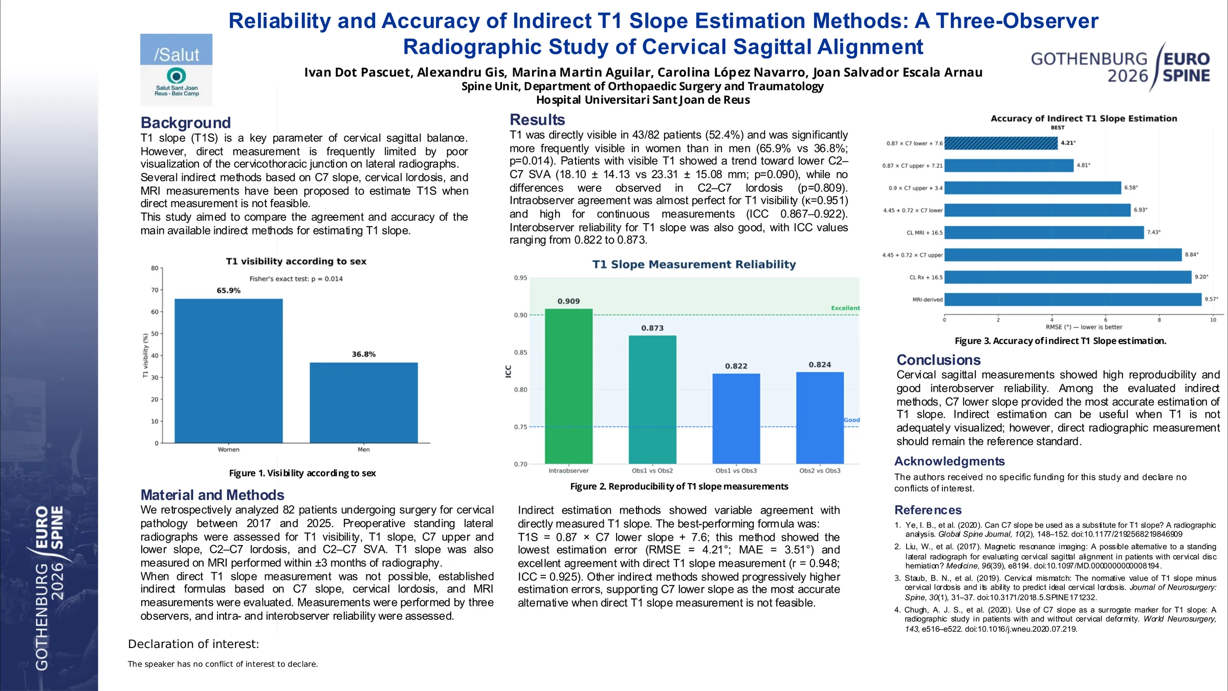
Eighty-two patients were included (44 women, 38 men). T1 visibility in measurement 1 was higher in women than in men (65.9% vs 36.8%; Fisher p=0.014). Patients with visible T1 showed a trend toward lower C2–C7 SVA (18.10 ± 14.13 vs 23.31 ± 15.08 mm), without statistical significance (p=0.090), and no differences were observed in cervical lordosis (p=0.809). Logistic regression did not identify SVA or lordosis as independent predictors of T1 visibility. Intraobserver agreement was almost perfect for T1 visibility (kappa=0.951), with high reliability for continuous measurements (ICC(3,1): 0.909 for T1 slope, 0.922 for C7 upper slope, 0.910 for C7 lower slope, and 0.867 for MRI T1 slope). Interobserver reliability remained good, with ICC(2,1) values ranging from 0.822 to 0.873. Among indirect estimation methods, T1 slope = 0.87 × C7 lower slope + 7.6 showed the best performance (RMSE=4.21; R²=0.899, n=41).
Cervical sagittal measurements demonstrated high intraobserver reproducibility and good interobserver agreement. Indirect formulas showed variable accuracy, with some providing close approximations but none fully replacing direct radiographic measurement. Indirect estimation may be useful when T1 visualization is limited, while direct T1 slope measurement remains the reference standard.