
Growing Spine
Lumbar Muscle Morphology and Composition in Adolescent Idiopathic Scoliosis: A Magnetic Resonance Study
- Bio-Medico Campus University Hospital, Rome, Italy
- Bambino Gesù Children's Hospital, Rom, Italy
- Agostino Gemelli University Policlinic, Roma, Italy
- Bambino Gesù Children's Hospital, Roma, Italy
Abstract
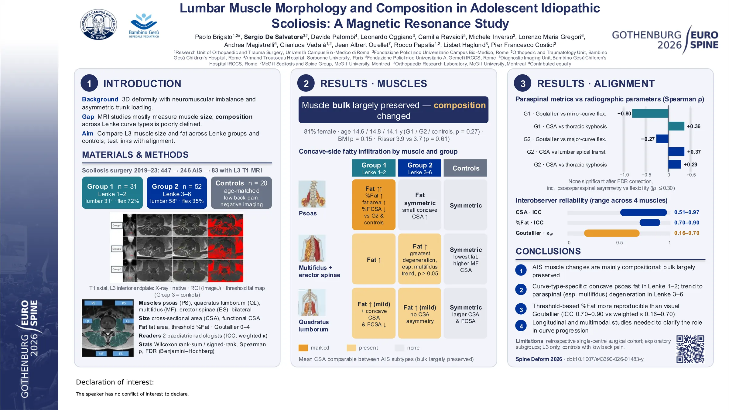
Adolescent idiopathic scoliosis (AIS) is a heterogeneous three-dimensional spinal deformity associated with neuromuscular imbalance and chronic asymmetric loading, which may induce adaptive changes in lumbar paraspinal and trunk muscles. Magnetic resonance imaging allows in vivo assessment of muscle morphology, most studies have focused on muscle size, with limited evaluation of muscle composition and few analyses across Lenke curve types. This study aimed to characterize lumbar muscle morphology and composition in AIS and to assess curve-type–specific and concave–convex asymmetry patterns.
This retrospective cohort study included AIS patients undergoing posterior spinal fusion between January 2019 and December 2023 with available preoperative T1-weighted axial MRI. Lumbar muscle morphology of the psoas, paraspinal (erector spinae and multifidus), and quadratus lumborum muscles was assessed bilaterally at the inferior endplate of L3. Patients were stratified by Lenke classification into non-structural lumbar curves (Group 1, Lenke 1–2) and structural lumbar curves (Group 2, Lenke 3–6) and compared with age-matched controls (Group 3). Muscle size and composition were quantified using threshold-based methods, with measurements independently performed by two pediatric radiologists and interobserver reliability assessed using intraclass correlation coefficients. Non-parametric analyses were performed using Wilcoxon tests for group comparisons and univariate Spearman correlations for associations between lumbar muscle metrics and radiographic parameters, with statistical significance set at p < 0.05.
Eighty-three AIS patients (Group 1, n = 31; Group 2, n = 52) and 20 controls were included. Demographic characteristics were comparable between groups. Lumbar muscle bulk was largely preserved across AIS subtypes and controls, with minimal cross-sectional area asymmetry. In contrast, muscle composition showed distinct curve-type–specific patterns. Group 1 demonstrated marked concave-sided fatty infiltration of the psoas muscle, whereas Group 2 exhibited greater degeneration of paraspinal muscles, particularly the multifidus (p>0.05). Controls showed lower fatty infiltration and no side-to-side asymmetry. Quadratus lumborum morphology was relatively preserved, with modest concave-sided alterations in AIS. Associations between AIS muscle parameters, curve severity, flexibility, and spinal alignment were weak and not statistically significant after correction (p>0.05).
Lumbar muscle alterations in AIS predominantly involve muscle composition rather than muscle bulk and follow curve-type–specific patterns consistent with localized adaptations to asymmetric loading. These findings support further longitudinal and multimodal studies to clarify the role of muscle composition in the pathophysiology and progression of spinal deformity.
Figures and tables
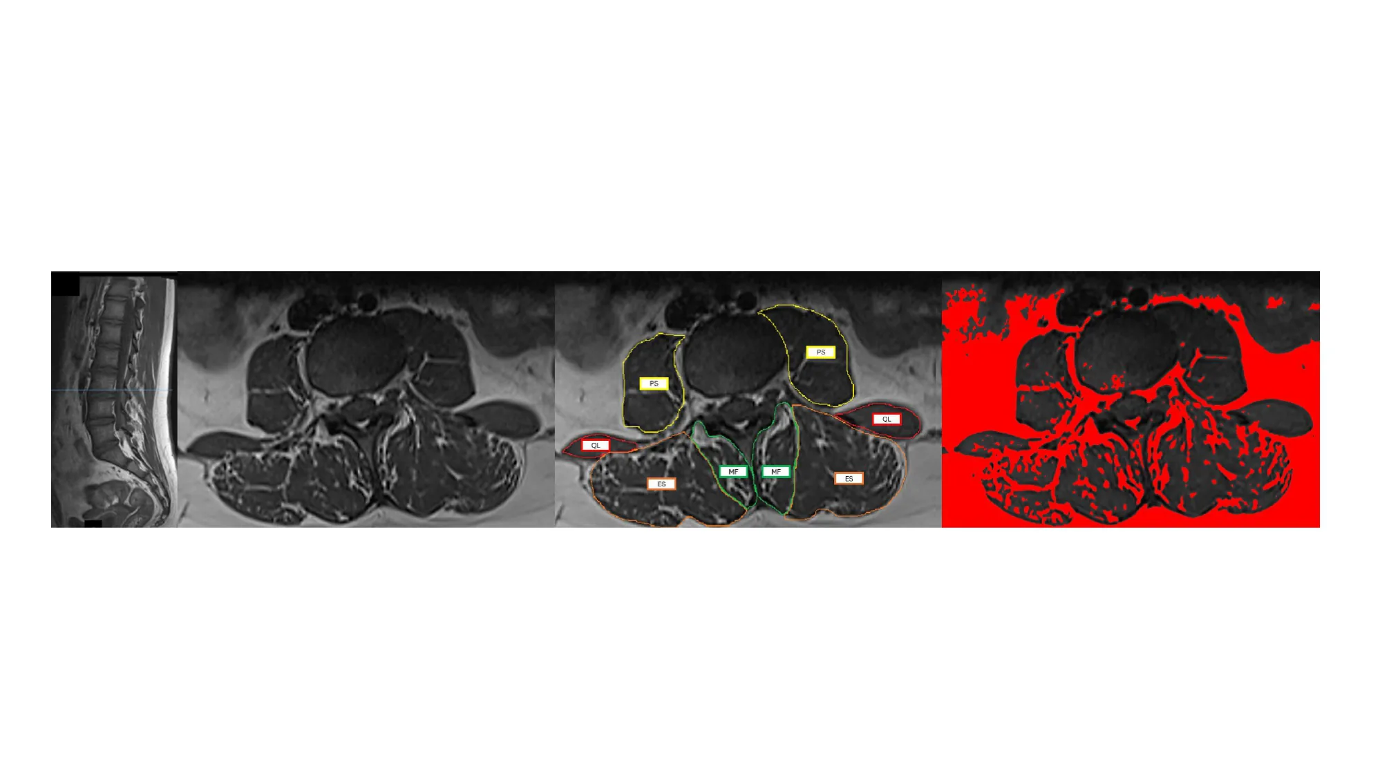
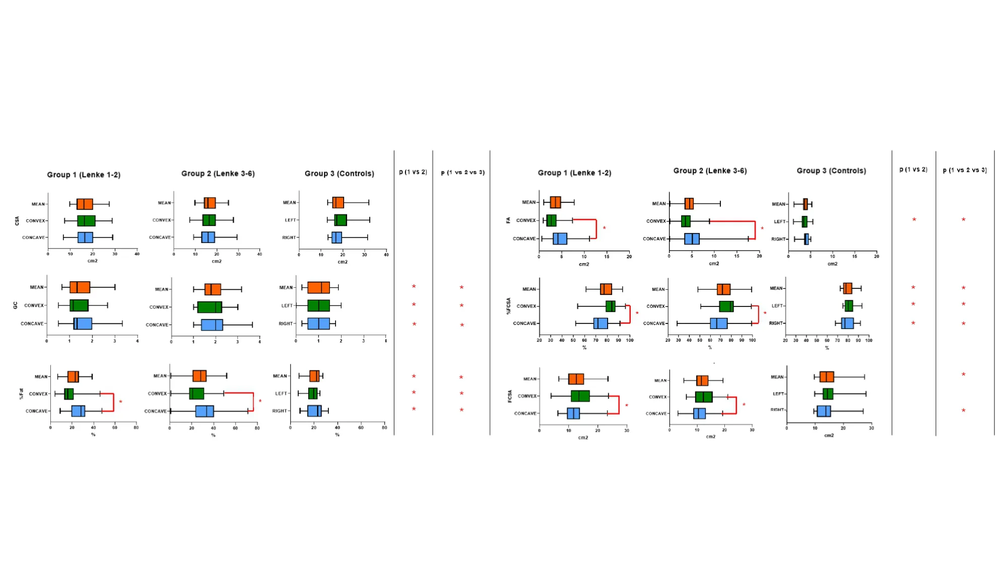
As submitted with the abstract. Tap a figure to open it at full size.