
Tumours & Infection
Endoscopic-Assisted Anterior Cervical Odontoidectomy with Preservation of the Atlas Anterior Arch for Odontoid Neoplasms
- The First Affiliated Hospital of Guangxi Medical University, Nanning, China
Abstract
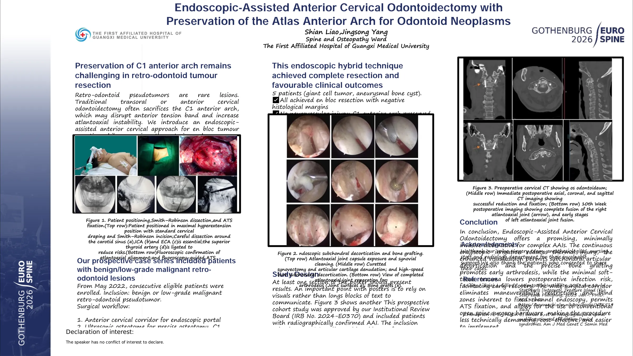
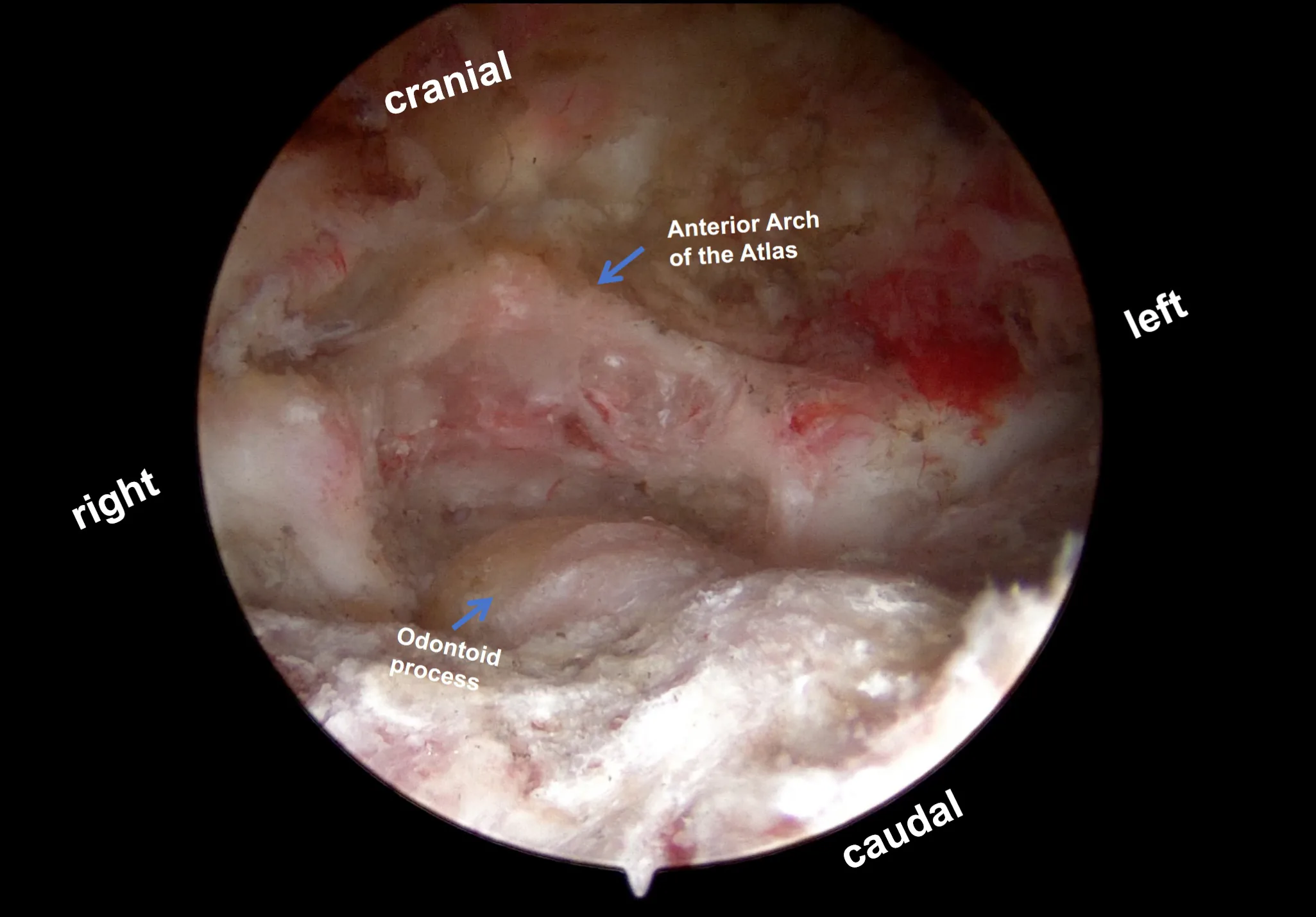
Odontoidectomy presents formidable challenges in spinal oncology. Conventional posterior approaches lack direct access for en bloc resection, while anterior transoral routes necessitate sacrifice of the anterior arch of the atlas, compromising the anterior tension band and carrying significant infectious risks. The transcervical approach offers a limited visual field and confined working space. This study describes a novel hybrid technique: an endoscopic-assisted anterior transcervical approach utilizing an ultrasonic osteotome to achieve en bloc odontoid resection with preservation of the atlas anterior arch, followed by immediate reconstruction via atlantoaxial lateral mass bone grafting and cannulated screw arthrodesis.
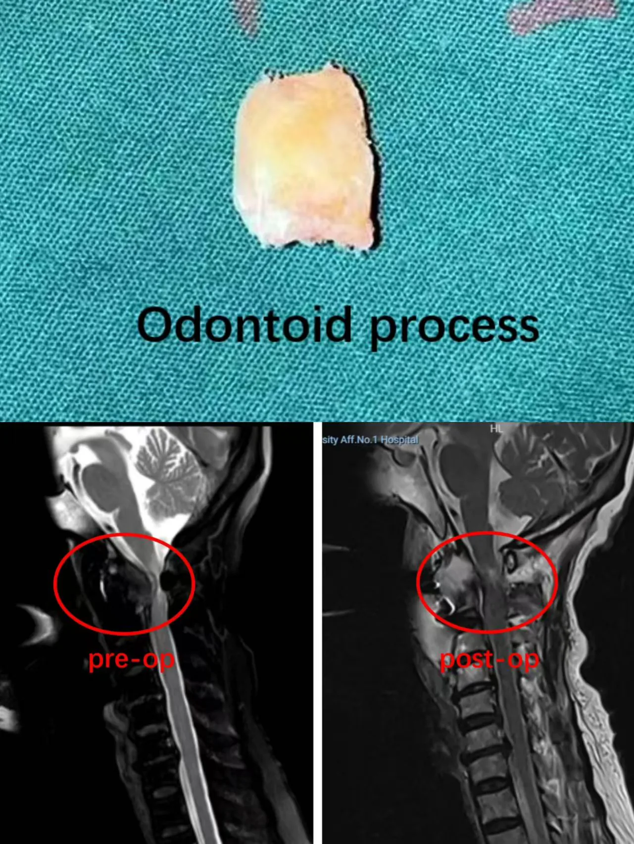
Since May 2022, patients diagnosed with odontoid tumors were prospectively enrolled. A standard anterior cervical approach was used to establish a spinal endoscopic working portal. Under the endoscopic view, meticulous dissection was carried down to the anterior aspect of the C2 vertebra. An ultrasonic osteotome was employed to perform precise osteotomy beyond the the odontoid process or tumor margins, achieving en bloc resection of the odontoid process. Subsequently, under combined endoscopic and fluoroscopic guidance, the articular surfaces of the atlantoaxial lateral joints were debrided, decorticated, and grafted with autologous iliac crest bone. Finally, percutaneous cannulated lag screws were placed to achieve compressive fixation across the lateral joints, providing immediate stability and promoting long-term fusion.
As of January 2026, five patients with odontoid tumors underwent the described procedure. All cases successfully underwent endoscopic en bloc odontoid resection with preservation of the anterior arch and primary fusion fixation. All instrumentation was accurately placed within the lateral masses. There were no intraoperative complications such as vertebral artery injury, dural violation, or neurological deficit. The mean operative time was 290 ± 50 minutes with an estimated blood loss of 400 ± 150 mL. No major postoperative complications occurred. At a mean clinical and radiological follow-up of 11 months, all patients demonstrated maintained or improved neurological status , significant reduction in axial pain , and radiographic evidence of progressive lateral mass fusion without implant failure or local tumor recurrence.
This surgical technique is a feasible and safe innovative strategy for treating odontoid tumors. Its offers a new paradigm for the minimally invasive surgical management of odontoid tumors and provides a new treatment option for eligible patients.
Figures and tables
As submitted with the abstract. Tap a figure to open it at full size.