
Basic Science & Economics
Reliability and accuracy of endplate angle measurement in lateral lumbar radiographs: A validation study using Digitally Reconstructed Radiographs
- Schulthess Klinik, Zürich, Switzerland
Abstract
Accurate measurement of lumbar endplate angles is essential for calculating sagittal spinal alignment parameters used in surgical planning. These measurements are routinely performed on PACS workstations using landmark-based tools, yet no empirical evidence exists to guide the choice of landmark selection strategy. This study compared the reliability and accuracy of three intuitive strategies for measuring endplate angles on lateral lumbar radiographic projections.
Fifty lumbar CT scans from a public dataset were used to generate Digitally Reconstructed Radiographs (DRRs) simulating standard and rotated (±10° axial and coronal) projections. Three-dimensional endplate angles at various lumbar levels (L1, L4, L5, S1) were established as ground truth via least-squares plane fitting to CT landmarks. Three raters measured endplate angles using three strategies: Extreme Points (anterior/posterior corners only), Peripheral Exclusion (medial points avoiding osteophytes), and Best Fit (least-squares regression through four landmarks). Intraclass correlation coefficients (ICC), mean absolute error (MAE), and rotation sensitivity were assessed.
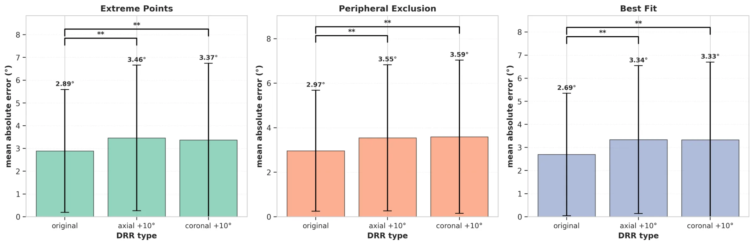
Best Fit achieved the highest inter-rater reliability (ICC = 0.97) and lowest mean absolute error (MAE) (3.1°), followed by Extreme Points (ICC = 0.95, MAE = 3.2°) and Peripheral Exclusion (ICC = 0.87, MAE = 3.4°). All techniques showed excellent intra-rater reliability (ICC > 0.95) and strong correlation with ground truth (r > 0.96). Measurement error increased by ~20% with patient rotation and was 2–3 times higher at the lumbosacral junction than at mid-lumbar levels.
Best Fit and Extreme Points strategies outperform Peripheral Exclusion in both reliability and accuracy. The minimal detectable change of 3–7° implies that clinicians should exercise caution when interpreting small changes in sagittal alignment, particularly at L5–S1. DRRs represent a valuable methodological tool for validating radiographic measurement techniques.
Figures and tables
As submitted with the abstract. Tap a figure to open it at full size.