
Cervical & Neural Pathologies
Point-Of-Care Automated Imaging Tool for Early Degenerative Cervical Myelopathy
- The University Of Oklahoma Health Sciences Center, Oklahoma City, United States of America
- University of Oklahoma, Oklahoma City, United States of America
Abstract
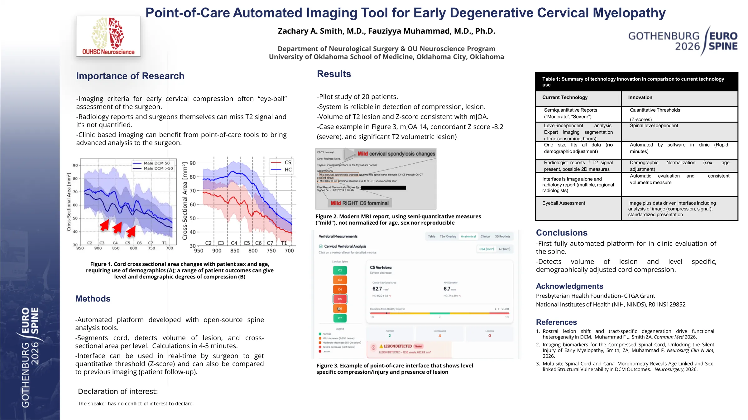
Degenerative cervical myelopathy (DCM) management relies on qualitative MRI interpretation, yet subclinical spinal cord compression is frequently undetected until irreversible neurological decline occurs. Quantitative morphometrics offer objective detection but remain inaccessible in clinical practice due to technical complexity. No fully automated system currently exists that translates raw T2-weighted (T2w) MRI into actionable quantitative reports for spine surgeons without neuroimaging expertise. We developed and validated such a platform, and evaluated its ability to identify subclinical compression and predict clinical trajectory through automated concordance analysis across the DCM spectrum.
A fully automated platform integrating validated open-source spinal cord analysis algorithms was developed. Clinicians upload T2w cervical MRI and mJOA scores through the interface. The system performs spinal cord and canal segmentation, vertebral labeling (C2-C7), intramedullary lesion detection, and per-level extraction of CSA, AP diameter, and canal dimensions. Metrics are normalized to a healthy control cohort (n=30) using Z-scores and C2-normalized ratios. A composite imaging severity score (0-10) integrating compression severity, canal compromise, and lesion burden is generated alongside automated clinical-imaging concordance classification (concordant, discordant, indeterminate). Twenty consecutive cervical T2w MRI datasets were processed to assess diagnostic yield and processing efficiency.
All 20 datasets achieved complete vertebral identification (C2-C7) within 4-6 minutes. Three cases illustrate the platform's clinical utility across the DCM spectrum. Case 1 (mJOA 18/18, neurologically intact): the system detected occult focal C5 compression (CSA 62.7 mm2, Z-score -2.30, CSA/C2 ratio 0.884, composite 4/10), classifying the case as discordant. This compression was not identified on qualitative radiological review. Case 2 (longitudinal, mJOA 18/18 at baseline): severe multilevel atrophy (composite 9/10) with a 632.8 mm3 intramedullary lesion was quantified despite normal function. Follow-up demonstrated deterioration to mJOA 14/18, confirming the predictive value of quantitative discordance for clinical surveillance. Case 3 (mJOA 9/18, severe DCM): concordance was confirmed with critical C4 compression (CSA 26.99 mm2, Z-score -8.2, lesion volume 285.7 mm2), validating the scoring framework against established presentation.
This is a novel, fully automated platform capable of generating quantitative spinal cord morphometry reports from standard clinical T2w MRI without specialist expertise. The system detected subclinical compression missed by qualitative assessment and demonstrated predictive value for clinical deterioration through concordance analysis. This directly addresses AO Spine RECODE-DCM priorities for standardized imaging criteria.
Figures and tables
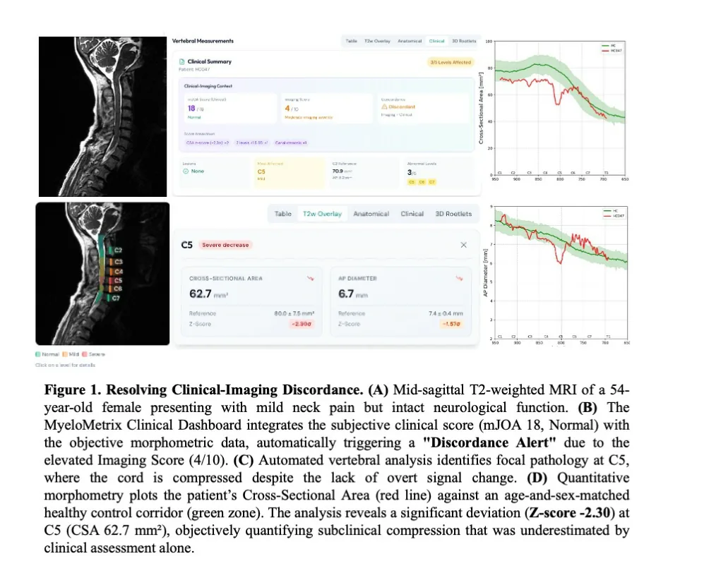
As submitted with the abstract. Tap a figure to open it at full size.