
Adult Deformity
Pedicle Subtraction Osteotomy in Lumbo-Sacral Fusion Surgery: Radiographic Evaluation of Lumbar Lordosis Behavior with Pre-Bent Rods
- Institut de la Colonne Vertébrale Lyon Charcot, Sainte-Foy-lès-Lyon, France
- Université Claude Bernard Lyon 1, Villeurbanne, France
Abstract
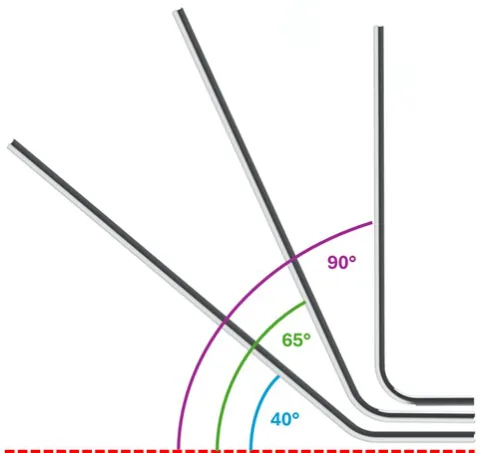
Sagittal balance restoration is a key objective in adult spinal deformity surgery. Pedicle subtraction osteotomies (PSOs) at L4 or L5 are sometimes performed with 40°, 65°, or 90° pre-contoured rods, but the specific impact of these rod angulations on achieved lumbar lordosis has not been systematically evaluated. The main objective is to assess the biomechanical effect of pre-bent rods on postoperative lumbar lordosis following lumbosacral fusion with PSO, and to identify preoperative spinopelvic predictors of lordosis differences between rod and lumbar spine.
Fifty-three patients (mean age 52 ± 16 years) who underwent L4 or L5 PSO with lumbosacral fusion and fixation (levels of spinal arthrodesis varied and could extend up to T2) using 40°, 65°, or 90° pre-contoured titanium rods were enrolled in this study. Spinopelvic parameters were measured on calibrated EOS imaging: pelvic incidence (PI), pelvic tilt (PT), sacral slope (SS), global lumbar lordosis (Global LL), L1–S1 lordosis (L1–S1 LL), and spinal lordosis ratio (SLR). Postoperative alignment was also assessed by computing lordosis differences between rod angulations and Global LL (ΔL Global), and lordosis differences between rod angulation and L1–S1 LL (ΔL L1–S1). Preoperative and postoperative EOS radiographs were analyzed with KEOPS software. Paired and repeated measures ANOVA assessed changes in spinopelvic parameters. Group comparisons were performed by sex, revision status, osteotomy level, rod curvature, and fusion length. Correlations between ΔL Global, ΔL L1–S1 and preoperative parameters were evaluated with Pearson coefficients.
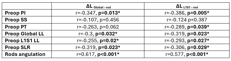
Global LL increased by 6.25° (11.56%, p=0.007) and L1–S1 LL by 9.78° (20.06%, p<0.001). SLR improved by 92.68% (p=0.05), reflecting focal correction at the lumbosacral junction. However, postoperative lordosis was systematically lower than rod angulation, with mean mismatches of –11.3° (Global LL) and –13.1° (L1–S1 LL). No significant differences in lordosis differences (ΔL Global and ΔL L1–S1) were observed across surgical subgroups. Higher preoperative PI, PT, Global LL and L1–S1 LL correlated with smaller ΔL Global and ΔL L1–S1 values, meaning that in patients with greater sagittal imbalance, the postoperative lumbar lordosis achieved after PSO tended to approximate more closely the intended curvature of the pre-contoured rod, with less mismatch between planned and actual correction.
PSOs at L4 or L5 combined with pre-contoured rods effectively restore lumbar lordosis and redistribute sagittal alignment. However, persistent mismatch between rod curvature and postoperative alignment underscores the influence of patient-specific anatomy and the need for individualized correction strategies.
Figures and tables
As submitted with the abstract. Tap a figure to open it at full size.