
Growing Spine
Pediatric Posterior Atlantoaxial Transarticular Screws Require a More Lateral Trajectory Due to Age-Related Atlantoaxial Morphology
- Yokohama Rosai Hospital, Yokohama, Japan
Abstract
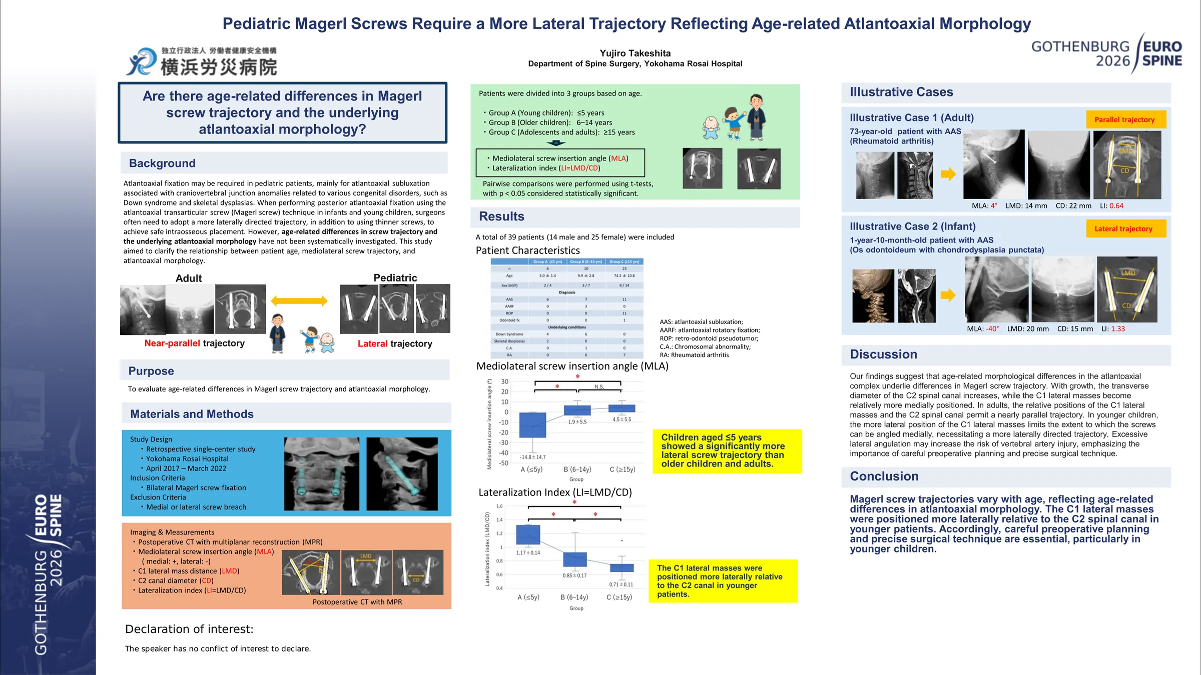
Atlantoaxial fixation may be required in pediatric patients, mainly for atlantoaxial subluxation associated with craniovertebral junction anomalies related to various congenital disorders, such as Down syndrome and skeletal dysplasias. When performing posterior atlantoaxial fixation using the atlantoaxial transarticular screw (Magerl screw) technique in infants and young children, surgeons often need to adopt a more laterally directed trajectory, in addition to using thinner screws, to achieve safe intraosseous placement. However, age-related differences in screw trajectory and the underlying atlantoaxial morphology have not been systematically investigated. This study aimed to clarify the relationship between patient age, mediolateral screw trajectory, and atlantoaxial morphology.
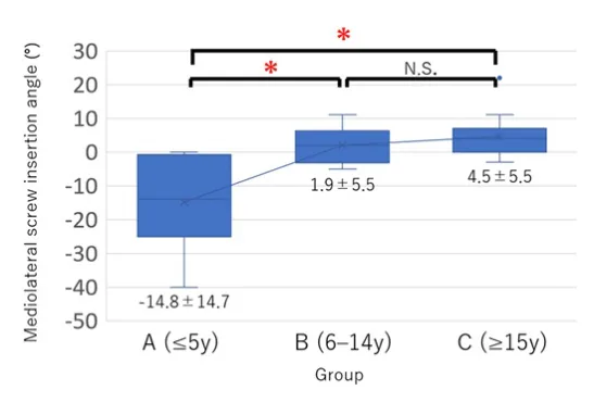
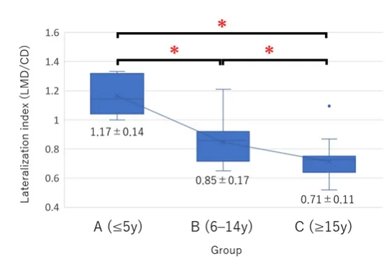
We retrospectively reviewed patients who underwent bilateral Magerl screw fixation at our institution between 2017 and 2022. Cases with any medial or lateral screw breach were excluded. Postoperative CT images were reconstructed to include both screws, and the following parameters were measured: mediolateral screw insertion angle (defined as the angle formed by bilateral screws; medial direction positive, lateral negative), lateral mass distance of the atlas (LMD), and spinal canal diameter of the axis (CD). The lateralization index (LMD/CD) was calculated to represent the relative lateral position of the atlas. Patients were divided into three groups based on age at surgery: Group A (≤5 years), Group B (6–14 years), and Group C (≥15 years). Mediolateral screw insertion angles and lateralization indices were statistically compared among groups using the t-test (p < 0.05).
Thirty-nine patients were included: 6 in Group A (mean age 3.0 ± 1.4 years), 10 in Group B (9.9 ± 2.8 years), and 23 in Group C (74.2 ± 10.8 years). Mean mediolateral screw insertion angles were −14.8 ± 14.7°, 1.9 ± 5.5°, and 4.5 ± 5.5° for Groups A, B, and C, respectively. The lateralization index was 1.17 ± 0.14 in Group A, 0.85 ± 0.17 in Group B, and 0.71 ± 0.11 in Group C. Younger patients demonstrated significantly more laterally directed screw trajectories and greater lateralization indices.
In younger patients, the atlas lateral mass is positioned more laterally relative to the axis spinal canal, necessitating a more laterally directed screw trajectory. These findings provide a morphological rationale for the laterally directed trajectory required in pediatric transarticular fixation. Recognition of this age-specific anatomy is critical to avoid vertebral artery injury and to achieve safe and accurate screw placement in young children.
Figures and tables
As submitted with the abstract. Tap a figure to open it at full size.