
Degenerative Thoracolumbar
Paraspinal Muscle Fatty Infiltration Is Associated With Nonunion and Cage Subsidence After Anterolateral Lumbar Interbody Fusion: A Level-Specific Analysis
- Taichung Veterans General Hospital, Taichung, Taiwan
Abstract
Anterolateral lumbar interbody fusion (LIF) surgery has been shown to be a successful treatment option for patients with degenerative lumbar disease. However, the effect of paraspinal muscle (PM) degeneration on clinical outcomes and risk factors for nonunion and cage subsidence (CS) in LIF remains unclear.
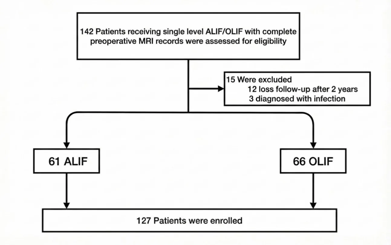
The study enrolled 127 patients who underwent ALIF or OLIF. Preoperative MRI was used to measure PM cross-sectional area (PMCSA) and the fatty infiltration (PMFI) at the superior and inferior levels of the fusion level. Fusion status and CS were assessed 2 years postoperatively.
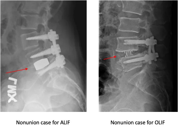
Smoking history, body mass index, and PMFI of the adjacent levels showed significant elevation in the nonunion and CS groups. PMFI at the inferior level emerged as a risk factor for nonunion and cage subsidence (p=0.026, odds ratio [OR]=1.12), particularly within the L1–L4 cohort. While multivariable analysis confirmed this association for L1–L4 levels, the correlation was less prominent in the L5–S1 subgroup. Nonunion of bone was observed when the PMFI rate surpassed 56.4% at the inferior level.
Qualitative muscle assessment is a more reliable predictor of fusion outcomes than quantitative size measurements, especially for L1–L4 levels. Preoperative PMFI evaluation should be integrated into surgical planning to improve patient selection and risk management.
Figures and tables
As submitted with the abstract. Tap a figure to open it at full size.