
Trauma
Metabolic Alterations and Sagittal Alignment in Spontaneous Osteoporotic Vertebral Compression Fractures: The Role of Vitamin D and Parathyroid Hormone
- Clinical Center of Vojvodina, Novi Sad, Serbia
Abstract
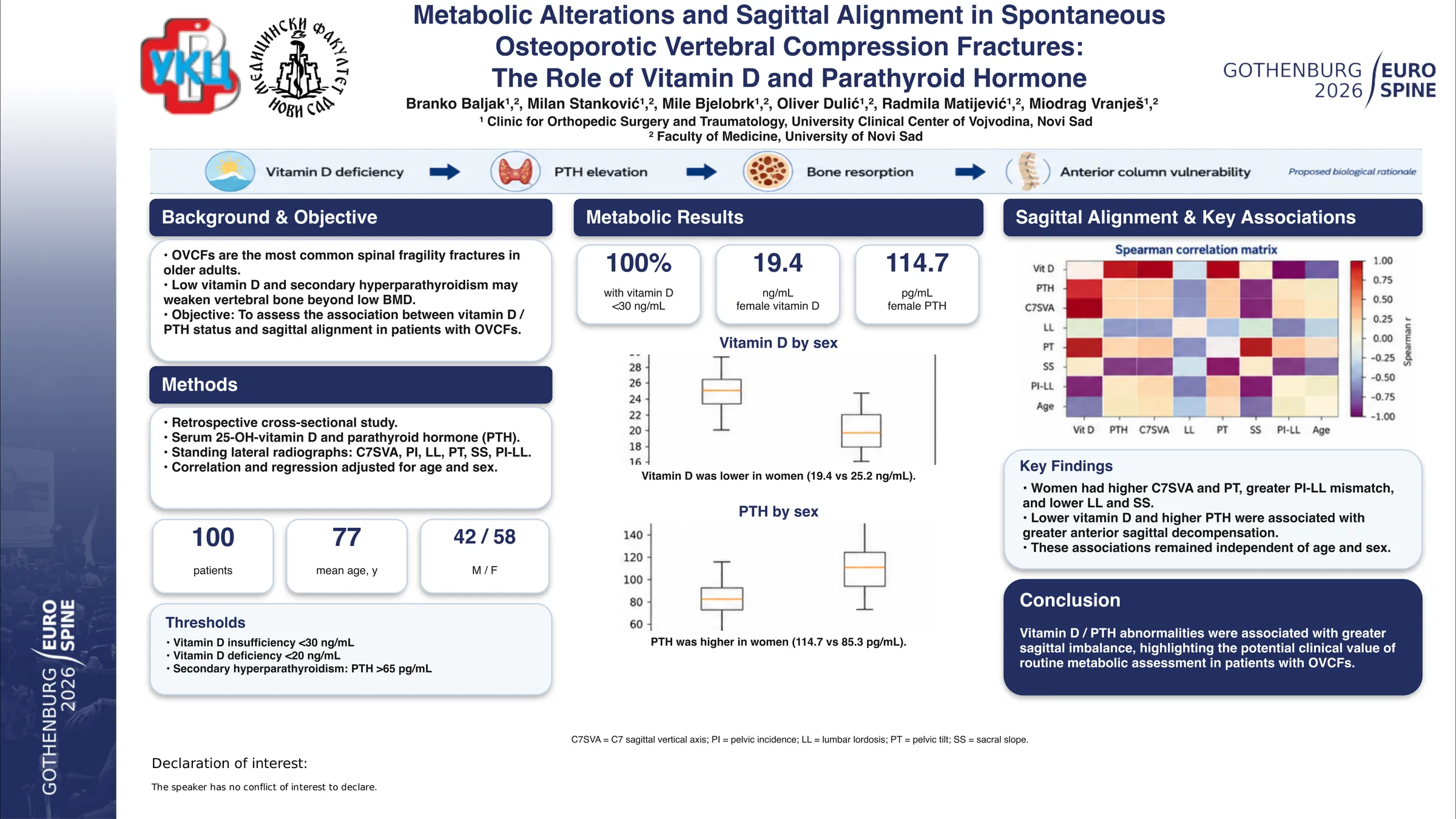
Spontaneous osteoporotic vertebral compression fractures (OVCFs) are incredibly common, frequently affecting older adults. They are, in fact, the most prevalent spinal fragility breaks. For ages, we've blamed these on low bone mineral density (BMD). Yet, increasing data points to something else: bone homeostasis disruptions. Things like vitamin D deficiency and secondary hyperparathyroidism play a big role, fundamentally weakening the vertebral body's inner structure. A lack of vitamin D, for example, causes parathyroid hormone (PTH) levels to spike. This, in turn, boosts osteoclastic activity and speeds up bone resorption. That sequence of events can seriously jeopardize the anterior spinal column, fostering a worsening forward curve and overall sagittal imbalance. Oddly, even with loads of research on metabolic factors in spinal fusion surgery, we still don't fully understand the metabolic makeup of spontaneous OVCFs in patients. Its connection to sagittal alignment? Still largely a mystery.
A retrospective cross-sectional study included 100 elderly patients (42 males, 58 females; mean age 77 years) with OVCFs. Patients without available serum 25-OH-vitamin D (VitD) and PTH measurements were excluded. Vitamin D insufficiency was defined as <30 ng/mL, deficiency as <20 ng/mL, and secondary hyperparathyroidism as PTH >65 pg/mL. Sagittal alignment was evaluated on standing full-spine lateral radiographs by measuring C7 sagittal vertical axis (C7SVA), pelvic incidence (PI), lumbar lordosis (LL), pelvic tilt (PT), sacral slope (SS), and PI–LL mismatch. Descriptive and comparative analyses were performed, and associations between metabolic parameters and sagittal alignment were assessed using correlation and regression models adjusted for age and sex.
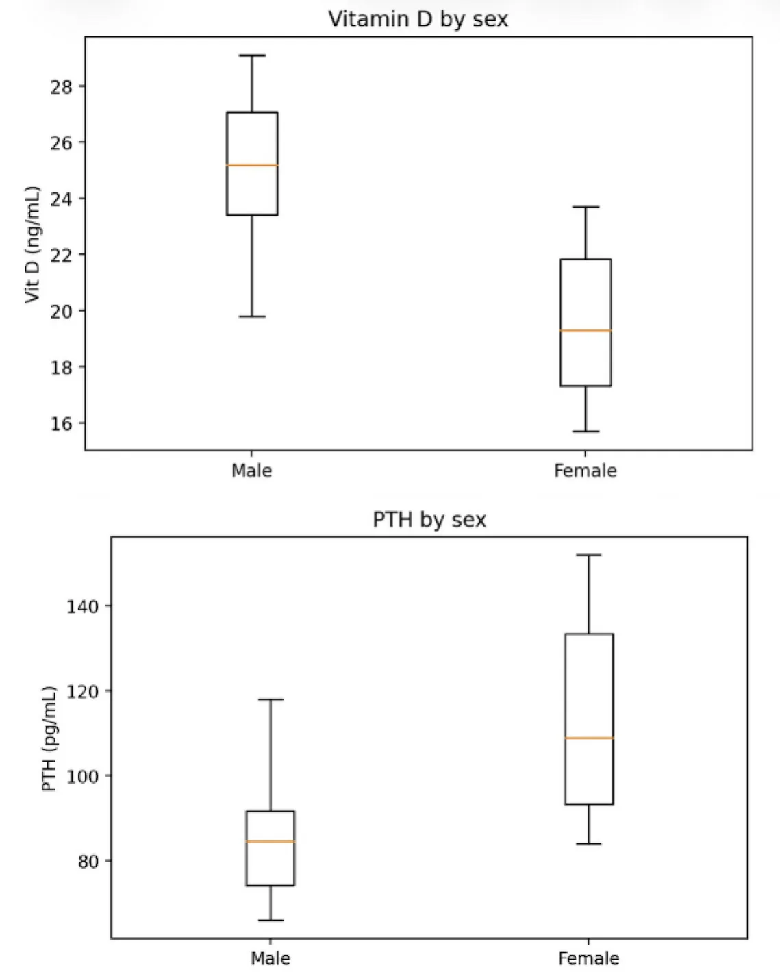
All 100 patients had vitamin D insufficiency, with deficiency predominantly in women. Females showed lower vitamin D (19.4 vs 25.2 ng/mL) and higher PTH levels (114.7 vs 85.3 pg/mL) compared to males.
Women demonstrated greater sagittal imbalance, including higher C7SVA, lower lumbar lordosis, higher pelvic tilt, lower sacral slope, and increased PI–LL mismatch.
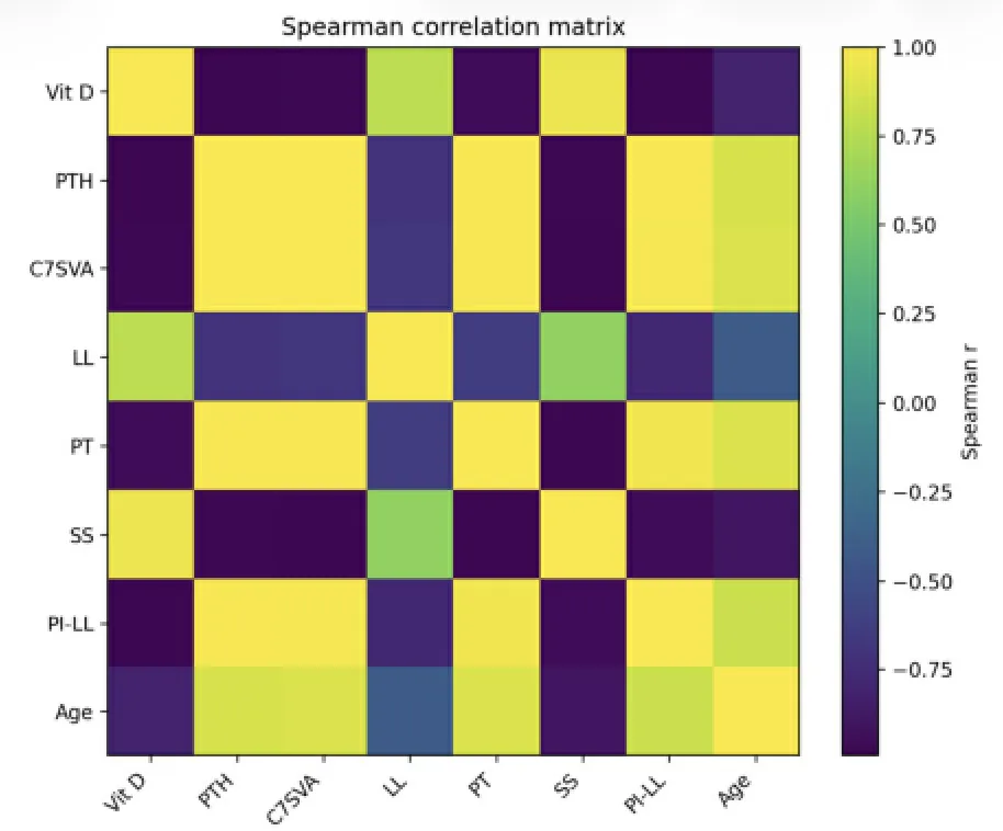
Lower vitamin D and elevated PTH were consistently associated with more pronounced anterior sagittal decompensation, independent of age and sex.
Vitamin D insufficiency and secondary hyperparathyroidism were highly prevalent in elderly patients with osteoporotic vertebral compression fractures, particularly among women, and were associated with greater sagittal imbalance. These findings suggest that impaired skeletal metabolic homeostasis may contribute to anterior sagittal decompensation, underscoring the importance of routine metabolic assessment in patients with OVCFs.
Figures and tables
As submitted with the abstract. Tap a figure to open it at full size.