
Degenerative Thoracolumbar
Location of stenosis matters: Unilateral Foraminal Lumbar Spinal Stenosis Is Associated with Asymmetric Paraspinal Muscle Degeneration
- Ulm University Medical Centre, Ulm, Germany
- University of Basel, Basel, Switzerland
- University Hospital Basel, Basel, Switzerland
Abstract
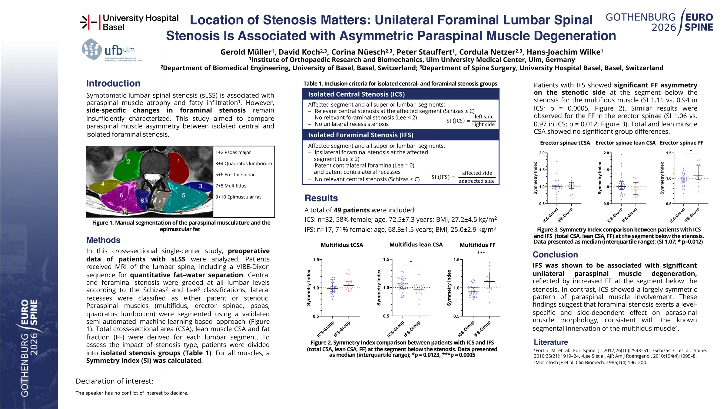
Symptomatic lumbar spinal stenosis (sLSS) is associated with paraspinal muscle atrophy and fatty infiltration. However, side-specific changes in foraminal stenosis remain insufficiently characterized. This study aimed to compare paraspinal muscle asymmetry between isolated central and isolated foraminal stenosis.
In this cross-sectional single-center study, embedded within a prospective umbrella study, preoperative data of patients with sLSS were analyzed. Patients received MRI of the lumbar spine, including a VIBE-Dixon sequence for quantitative fat–water separation. Central and foraminal stenosis were graded at all lumbar levels according to the Schizas and Lee classifications; lateral recesses were classified as either patent or stenotic. Paraspinal muscles (multifidus, erector spinae, psoas, quadratus lumborum) were segmented using a validated semi-automated machine-learning-based approach. Total cross-sectional area (CSA), lean muscle CSA and fat fraction (FF) were derived in each slice and averaged between the middle of the upper and lower intravertebral disc to receive one value for each lumbar segment.
To assess the impact of stenosis type, patients were divided into isolated central stenosis (ICS; central stenosis ≥ C (Schizas), no relevant foraminal stenosis) and isolated foraminal stenosis (IFS; affected segment and all superior segments ipsilateral foraminal stenosis ≥ 2 (Lee) but patent contralateral foramina (Lee, 0) and no relevant central stenosis (Schizas < C).For all muscles, a symmetry index (SI) was calculated. In the IFS group, the SI was defined as ratio of the affected (side with foraminal stenosis) to the unaffected side; in the ICS group as ratio of the left to the right side. Between-group comparisons were performed using independent-samples t-tests (p < 0.05).
A total of 48 patients were included (ICS: n = 31, 58% female; IFS n = 17, 71% female). Mean age was 72.5 ± 7.3 years in the ICS group and 68.3 ± 11.5 years in the IFS group. Mean BMI was 27.2 ± 4.5 and 25 ± 2.9 kg/m2, respectively.
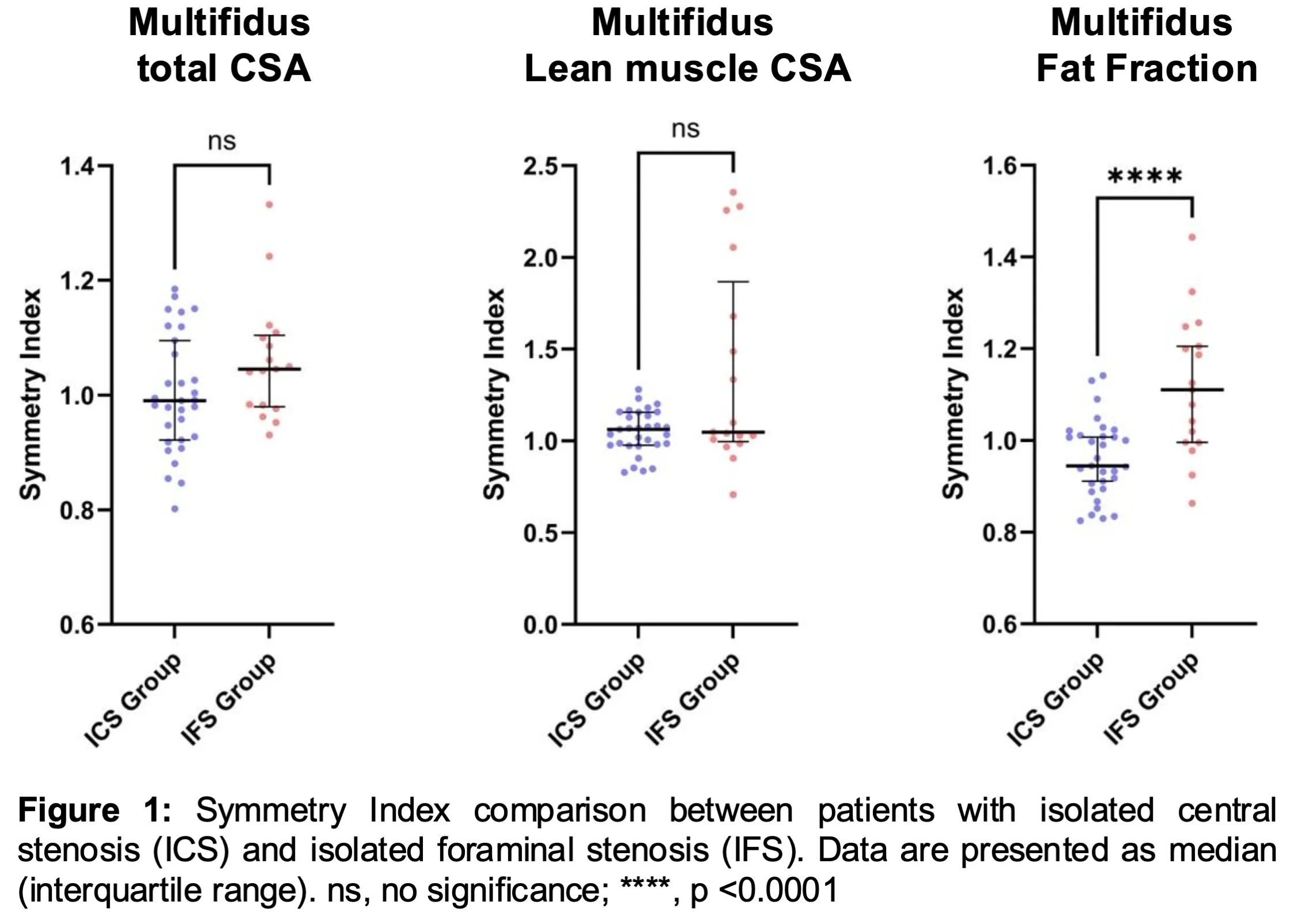
Patients with IFS showed significant FF asymmetry on the stenotic side at the segment below the stenosis for the multifidus muscle (SI 1.12 vs 0.96 in ICS; p < 0.001, Figure 1). Similar results were observed for the FF in the erector spinae (SI 1.09 vs 0.98 in ICS; p = 0.012). Total and lean muscle CSA showed no significant group differences.
IFS was associated with significant unilateral paraspinal muscle degeneration, reflected by increased FF at the segment below the stenosis. In contrast, ICS showed a largely symmetric pattern of paraspinal muscle involvement. These findings suggest that foraminal stenosis exerts a level-specific and side-dependent effect on paraspinal muscle morphology, consistent with the known segmental innervation of the multifidus muscle.
Figures and tables
As submitted with the abstract. Tap a figure to open it at full size.