
Adult Deformity
Is the Updated Roussouly Classification Truly Categorical? A Multicenter Analysis of 525 Asymptomatic Adults Identifying an Anteverted Transitional Sagittal Morphotype (Type 3–4AP)
- Etlik Şehir Hastanesi Ortopedi ve Nöroloji Hastanesi, Ankara, Türkiye
- Istanbul Science University, Istanbul, Türkiye
- Baltalimanı Kemik Hastalıkları Eğitim ve Araştırma Hastanesi, Levent Semt Polikliniği, Istanbul, Türkiye
- Gazi University Faculty of Medicine, Ankara, Türkiye
- Afyonkarahisar Sağlık Bilimleri Üniversitesi (AFSÜ), Afyonkarahisar, Türkiye
- Private, İzmir, Türkiye
- Balıkesir Atatürk City Hospital, Balıkesir, Türkiye
- Süleyman Demirel Üniversitesi Tıp Fakültesi Dekanlığı, Isparta, Türkiye
- Ondokuz Mayıs University, Samsun, Türkiye
- Anatolian Spine Study Group - ASSG, Istanbul, Türkiye
- Adnan Menderes University, Aydın, Türkiye
- Nya Karolinska, Stockholm, Sweden
Abstract
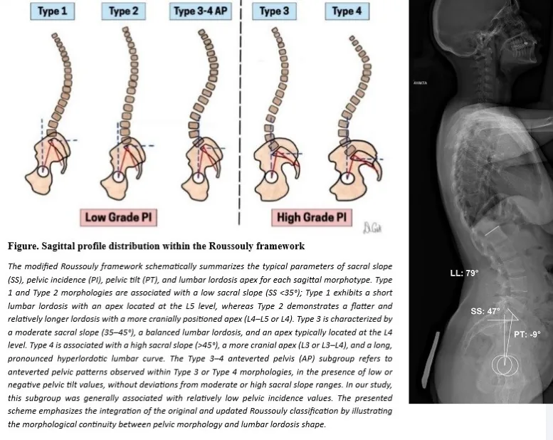
The Updated Roussouly Classification (URC) describes sagittal spinal alignment using four discrete morphotypes defined by pelvic incidence, sacral slope, and lumbar lordosis distribution. Although widely used in adult spinal deformity assessment and surgical planning, the strictly categorical structure of URC may oversimplify the continuum of sagittal morphology. This study aimed to evaluate URC distribution in a large asymptomatic adult population and to investigate whether anteverted pelvic configurations represent a transitional sagittal phenotype between Type 3 and Type 4.
A retrospective multicenter radiographic analysis was conducted including 525 asymptomatic adults from 10 centers. Standardized standing full-spine lateral radiographs were assessed. Sagittal spinopelvic parameters (PI, PT, SS, lumbar apex) were independently measured by two observers and classified according to URC criteria. Particular focus was given to anteverted pelvic patterns characterized by low or negative PT values combined with moderate-to-high SS. Distribution patterns were compared with contemporary international normative datasets.
Among 525 participants (mean age 35.1 years; 374F/151M), Type 3 was the most prevalent morphotype (31.2%), followed by Type 2 (22.4%), Type 4 (20.2%), and Type 1 (10.2%). An anteverted pelvic configuration was identified in 15.5% of Type 3 and 1.5% of Type 4 subjects, forming a pooled transitional Type 3–4AP subgroup (16.0%). This subgroup was characterized by moderate-to-high sacral slope, low or negative pelvic tilt, relatively lower pelvic incidence, and a lumbar apex distribution consistent with Type 3–4 morphology. Type 4 prevalence increased with age, whereas the Type 3–4AP phenotype was more common in younger and middle-aged individuals. Although overall distributions were comparable to international normative cohorts, the categorical URC framework did not fully capture the morphological continuum observed between Type 3 and Type 4.
In this large multicenter normative analysis, a distinct anteverted transitional sagittal morphotype spanning Type 3 and Type 4 was identified in 16% of asymptomatic adults. These findings support the concept that sagittal alignment exists along a morphological continuum rather than strictly discrete Updated Roussouly Classification categories. Recognition of transitional anteverted phenotypes may enhance sagittal classification frameworks and improve interpretation of alignment patterns in adult spinal deformity planning. Longitudinal studies are needed to clarify the biomechanical and clinical implications of this phenotype.
Figures and tables
As submitted with the abstract. Tap a figure to open it at full size.