
Adult Deformity
Intraoperative ultrasound visualization of paravertebral anatomy in the retroperitoneal space during Mini-Open OLIF and Lateral ALIF in lateral decubitus position
- Interdisciplinary Spine Center, Frankfurt, Germany
Abstract
Mini-open oblique lumbar interbody fusion (OLIF) and lateral anterior lumbar interbody fusion (ALIF) performed in the lateral decubitus position are established minimally invasive techniques for lumbar spine reconstruction. Despite their advantages, these approaches traverse the retroperitoneal space and carry a risk of potentially serious visceral and vascular complications. The objective of this study was to evaluate the usefulness of intraoperative ultrasound for enhancing anatomical orientation and surgical safety during mini-open OLIF and lateral ALIF procedures.
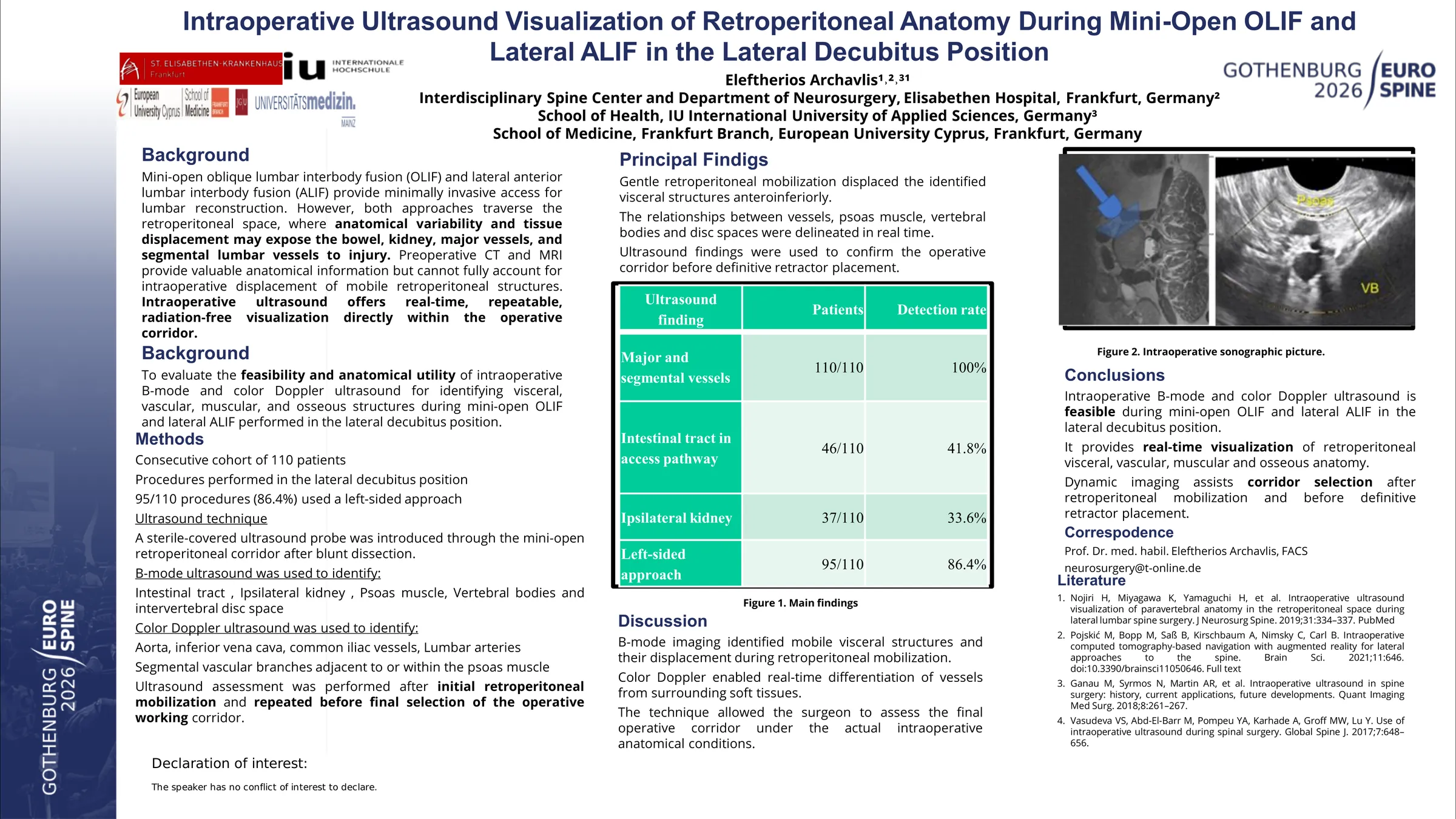
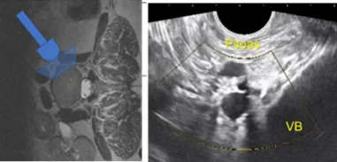
Intraoperative ultrasound was performed using a sterile-covered transvaginal ultrasound probe inserted into the retroperitoneal operative corridor during surgery in lateral position. B-mode ultrasound was used to identify the intestinal tract, kidneys, psoas muscle, and vertebral bodies. Color Doppler mode was applied to visualize the aorta, vena cava, common iliac vessels, lumbar arteries, and their segmental branches. Ultrasound assessment was conducted after blunt retroperitoneal dissection and during final confirmation of the safe working corridor.
The study cohort consisted of 110 patients undergoing mini-open OLIF or lateral ALIF, with 95 procedures performed via a left-sided approach. The intestinal tract and ipsilateral kidney along the surgical access pathway were visualized in 46 and 37 patients, respectively. Gentle retroperitoneal mobilization displaced the visceral structures anteroinferiorly, allowing confirmation of a safe corridor anterior to the psoas muscle. In all cases, major anterior vascular structures and segmental lumbar vessels within or adjacent to the psoas muscle were clearly identified using Doppler ultrasound. The spatial relationships between vessels, vertebral bodies, intervertebral discs, and the psoas muscle were reliably delineated in real time.
During mini-open OLIF and lateral ALIF performed in lateral decubitus position, intraoperative ultrasound enables real-time visualization of retroperitoneal visceral and vascular anatomy along the surgical access path. This technique enhances anatomical safety, facilitates precise corridor selection, and reduces the risk of visceral and vascular injury without adding invasiveness to the procedure.
Figures and tables
As submitted with the abstract. Tap a figure to open it at full size.