
Growing Spine
Impact of posterior spinal fusion on daily life activities in Lenke-1 AIS patients: a 3D functional evaluation
- Saint Joseph University of Beirut, Beirut, Lebanon
- Arts et Métiers, Paris, France
Abstract
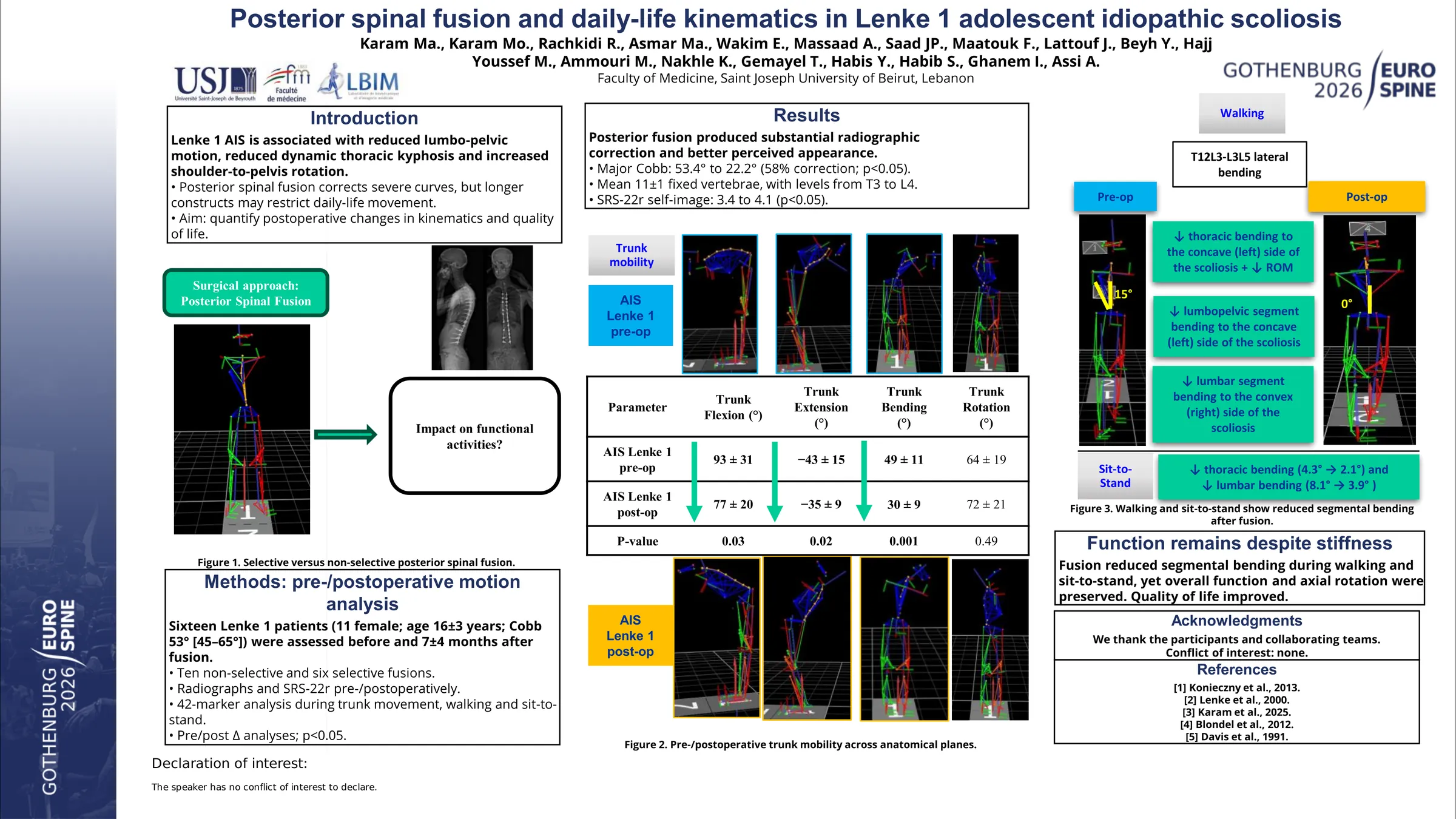
Various functional adaptations have been reported in daily life activities in non-operated AIS patients during walking, sit to stand and trunk anatomical movements, especially in the spinal segments. However, the impact of posterior spinal fusion (PSF) on these functional alterations is still unclear. The aim was to investigate full-body kinematic changes during daily life activities in AIS Lenke-1 patients following PSF.
16 AIS patients with Lenke-1 type (Cobb: 53±9°; 11F) scheduled for PSF using a concave rod translation combined with direct vertebral rotation were included, along with 21 healthy controls (ctr). All participants underwent standing biplanar radiographs and filled SRS-22r form. 3D movement analysis was performed during trunk anatomical movements (flexion, extension, bending and rotation), walking, and sit-to-stand (STS) for the control group and pre and post-operatively for the AIS group. The pre to postoperative variation (Δ=post-pre) of variables was calculated. Kinematic and radiographic parameters were compared to controls and pre vs post-operatively. Correlations between variables were analyzed.
Postoperatively, the average major Cobb was reduced to 22±8° ; the average number of fixed vertebrae was 11±1, ranging from T3 to L4. During anatomical trunk movements, Lenke-1 patients showed a postoperative reduction in the maximal trunk flexion (77 vs 93° in preop & 113° in ctr), extension (-35 vs -45° in preop & ctr), and lateral bending (-31 vs -48° in preop & ctr; all p<0.05). Those with longer fusion that included the lumbar spine (reaching L2 to L4) had more reduced sagittal ROM during trunk flexion (74 vs 90°, p=0.016). Trunk rotational range of motion (ROM) remained comparable to preoperative values and to ctr (68°). This was associated with a decreased shoulder rotation relative to pelvis (30 vs 50° in preop & ctr) and an increased pelvic rotation (54 vs 35° in preop & ctr; Figure 1). During both walking and STS movements, thoracic, lumbar and lumbo-pelvic bending that were limited preoperatively, reached normal values postoperatively. The number of fused vertebrae was negatively correlated to the variation in trunk and lumbar segment bending during all movements (ρ=-0.6 and -0.5 resp.; all p<0.05). Self-image increased postoperatively (4.5 vs 2.5 in preop, p=0.01).
This introductory study showed that trunk mobility was significantly reduced primarily in the sagittal and frontal planes, especially in patients with non-selective fusion.
However, axial rotation was relatively preserved, likely due to compensatory pelvic motion in the transverse plane. Despite these trunk movement limitations, walking and sit-to-stand functional tasks were not adversely affected, while self-image improved.
Figures and tables
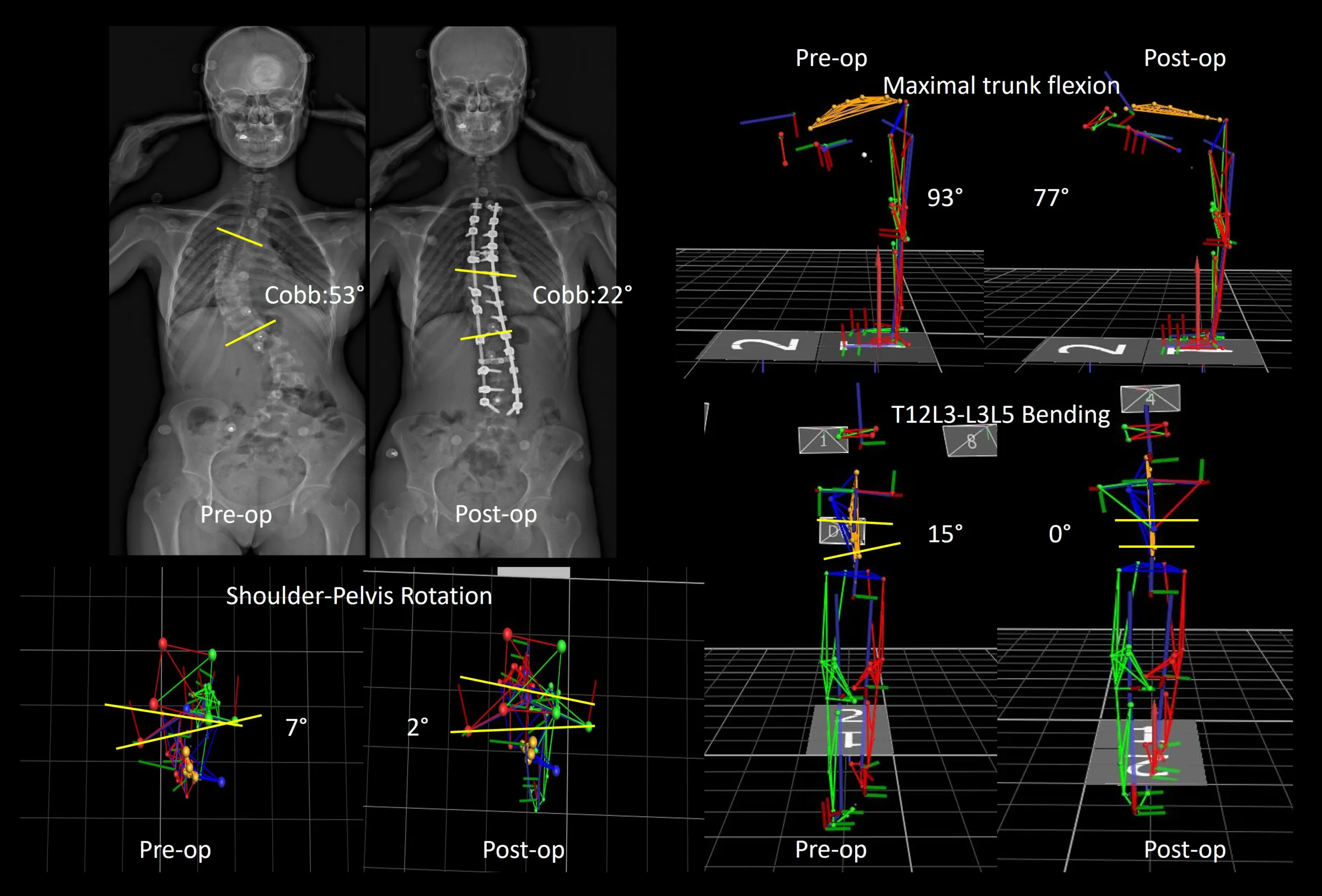
As submitted with the abstract. Tap a figure to open it at full size.