
Growing Spine
Immediate Biomechanical Effects of a 3D-Printed Scoliosis Brace on Coronal and Spinopelvic Alignment in Adolescent Idiopathic Scoliosis
- Hospital for Special Surgery Main Hospital, New York, United States of America
- Unimed Hospital, Belo Horizonte, Brazil
- Universidade Federal de Minas Gerais, Belo Horizonte, Brazil
Abstract
Three-dimensional (3D)-printed scoliosis braces enable patient-specific correction strategies for the treatment of adolescent idiopathic scoliosis (AIS). While coronal plane correction is expected with bracing, the immediate biomechanical effects on spinopelvic sagittal alignment are less well characterized. This study aim to quantify immediate coronal and sagittal radiographic alignment changes following application of a 3D-printed scoliosis brace in adolescent idiopathic scoliosis.
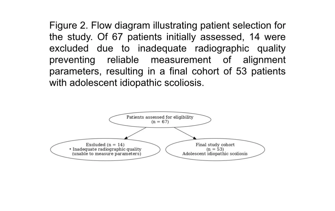
Fifty-three adolescents with adolescent idiopathic scoliosis treated with a 3D-printed brace were analyzed. Standing full-spine radiographs were obtained before and after brace application (in-brace). The primary coronal parameter was Cobb angle of the main curve, and the primary sagittal parameter was pelvic incidence–lumbar lordosis (PI–LL) mismatch. Secondary parameters included pelvic incidence (PI), lumbar lordosis (L1–S1), pelvic tilt (PT), sacral slope (SS), T1–pelvic angle (TPA), spinopelvic angle (SPA), spinosacral angle (SSA), and vertebral rotation (Nash–Moe). Pre- and post-corset values were compared using paired t-tests or Wilcoxon signed-rank tests as appropriate (α=0.05). As a secondary biomechanical analysis, PI–LL mismatch was categorized as balanced (−10° to +10°) or imbalanced, and transitions in sagittal alignment status were assessed using the McNemar test.
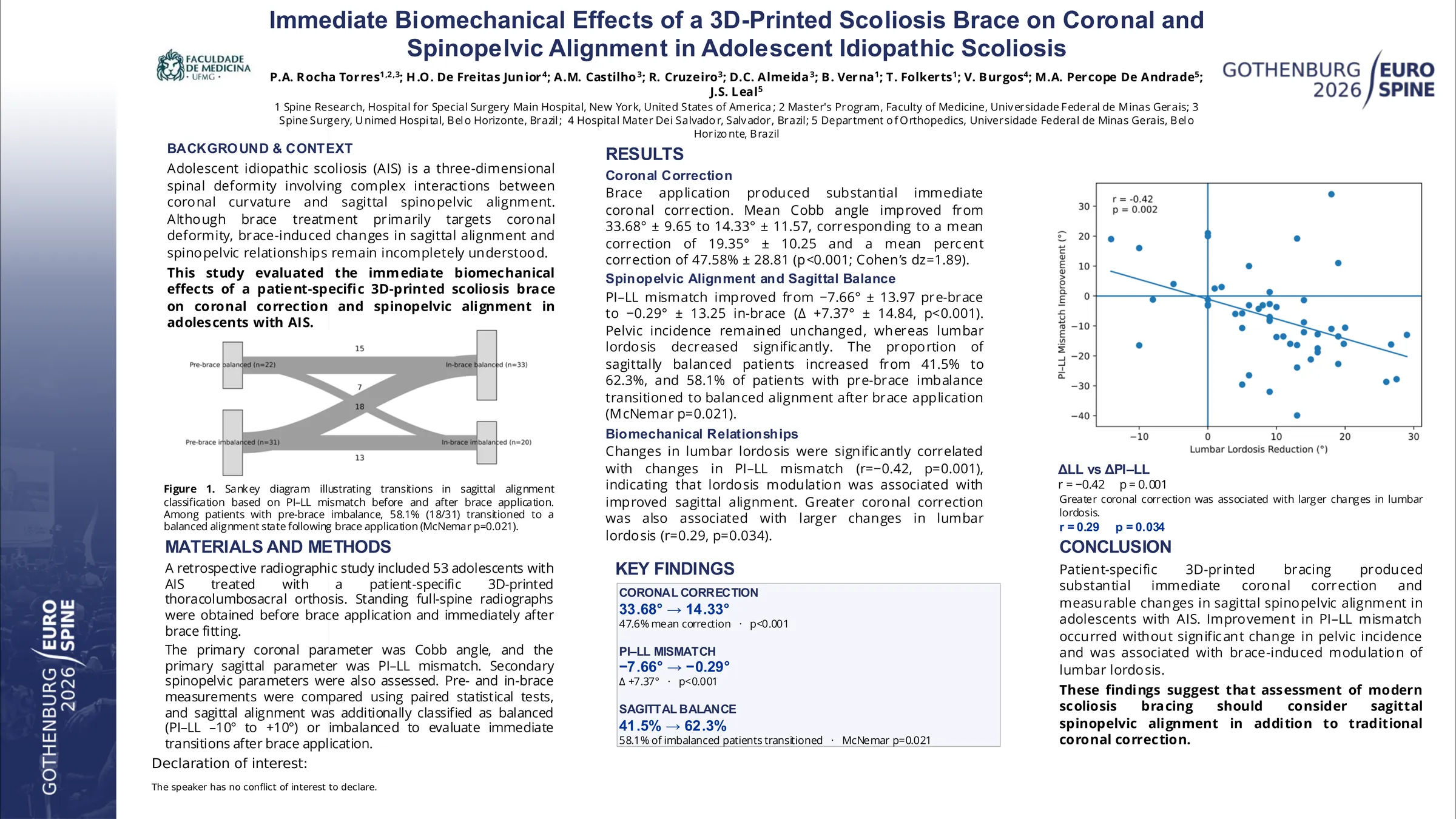
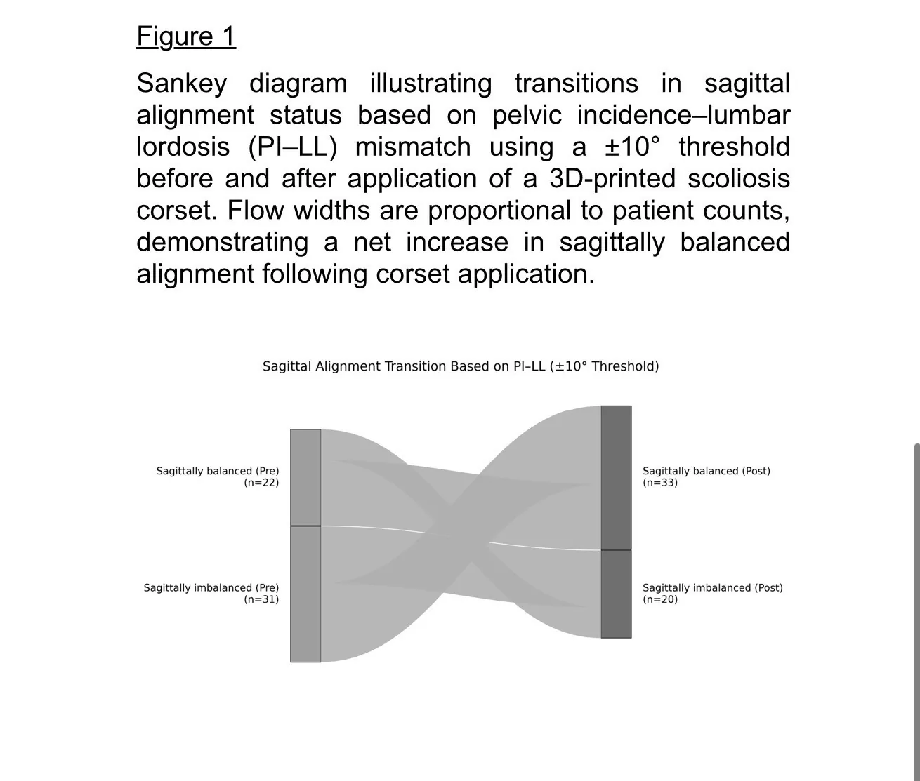
Mean Cobb angle improved from 33.68° ± 9.65 to 14.33° ± 11.57, representing a mean correction of 19.35° ± 10.25 (p<0.001; Cohen’s dz=1.89), with a mean percent correction of 47.58% ± 28.81. PI–LL mismatch improved from −7.66° ± 13.97 to −0.29° ± 13.25 (Δ +7.37° ± 14.84; p<0.001). Using a ±10° threshold, the proportion of sagittally balanced patients increased from 41.5% pre-brace to 62.3% post-brace; among patients with pre-brace PI–LL imbalance, 58.1% transitioned to a balanced alignment state (McNemar p=0.021). Pelvic incidence remained unchanged (p=0.521). Secondary sagittal parameters demonstrated modest but statistically significant changes.
In adolescent idiopathic scoliosis, application of a 3D-printed scoliosis brace resulted in large immediate coronal correction and significant biomechanical changes in spinopelvic alignment. These findings support that customized 3D-printed bracing can influence both coronal deformity and sagittal alignment parameters in-brace, warranting further investigation of biomechanical mechanisms and longer-term radiographic and clinical follow-up.
Figures and tables
As submitted with the abstract. Tap a figure to open it at full size.