
Growing Spine
Functional changes with age in idiopathic scoliotic patients: evaluation of kinematic limitations and their compensatory mechanisms
- Saint Joseph University of Beirut, Beirut, Lebanon
Abstract
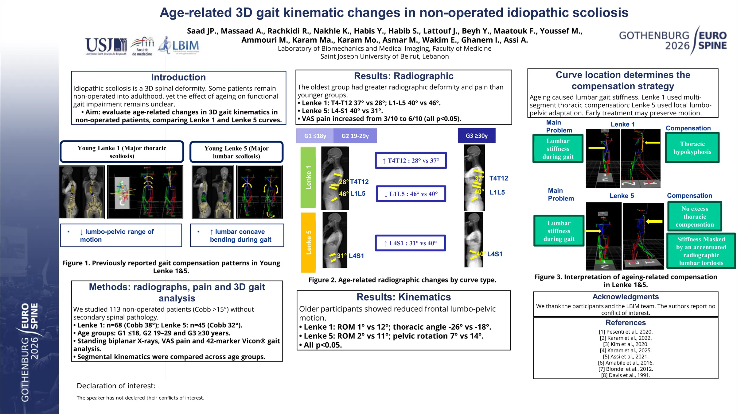
Scoliotic deformity affects self-image in adolescents and, if untreated, may progress to severe spinal deformity impairing quality of life and mobility. Although kinematic patterns across AIS curve types have been described, it remains unclear how non-operated scoliotic patients adapt their gait with ageing. This observational study compared 3D gait and compensatory mechanisms in scoliotic patients with the same curve type across age groups.
Non-operated adolescents and adults with idiopathic scoliosis underwent biplanar radiographs to extract spinopelvic parameters and 3D gait analysis to calculate full-body kinematics. Patients were classified by Lenke types and divided into 3 age groups: ≤18y (G1), 19-29y (G2), and ≥30y (G3). Spinopelvic parameters and kinematics were compared between groups and a normative database of 150 controls.
110 patients (11-70y) were included: 68 Lenke-1 (G1:40, G2:20, G3:8 patients; average Cobb: 38°) and 45 Lenke-5 (G1:23, G2:15, G3:7 patients; average Cobb: 32°). In Lenke 1 patients, the older group (G3) showed increased T4T12 (37 vs 28° in G1 & G2) and reduced L1L5 (40 vs 46°; both p<0.05) on static radiographs. Dynamically, young Lenke-1 patients (G1) had a thoracic concave bending (to the left) (T3T6-T6T9: -6 vs 0°) with compensatory lumbar bending (to the right) (T12L3-L3L5: 9 vs -2°) when compared to controls. These segmental alterations induced several kinematic adaptations: reduced lumbo-pelvic mobility (ROM L3L5-pelvis: 8 vs 12°), increased thoracic hypokyphosis (C7T9-T9T12: -18 in G1 vs 23°) with normal thoraco-lumbar hypolordosis (T9T12-T12L3: -4 in G1 vs -6°). G2 had similar patterns. In old Lenke-1 patients (G3), lumbo-pelvic frontal mobility further decreased (1 vs 12°) with an increased both thoracic hypokyphosis (-26 vs 23°) and thoraco-lumbar hypolordosis (12 vs -6°; all p<0.05). In Lenke-5 patients, the older group (G3) showed increased radiographic L4S1 angle (40 vs 31° in G1 & G2). Dynamically, young Lenke-5 patients(G1) had increased lumbar concave bending (T12L3-L3L5: 5 vs -2°) compared to controls. They maintained both normal pelvic axial mobility (14°) and frontal lumbo-pelvic mobility (T12L5-pelvis ROM: 11°). G2 had similar patterns. In old Lenke-5 patients (G3), there was a significant reduction in both pelvic axial mobility (7 vs 14°) and frontal lumbo-pelvic mobility (2 vs 11°;both p<0.05;Fig.1) compared to G1 & G2. Pain increased with age in both Lenke-1 and 5 (VAS:6 vs 3 in G1&G2,both p<0.05).
With age, Lenke-1 patients had increased kinematic alterations at the thoracic and lumbar levels. However, Lenke-5 patients had their kinematic alterations limited to the pelvis. Both Lenke-1 and 5 developed lumbo-pelvic rigidity with age, with increasing pain, suggesting progressive loss of compensatory capacity. This might be due to degenerative disc disease and facet joint arthritis that may play a role in kinematic alterations in ageing population.
Figures and tables
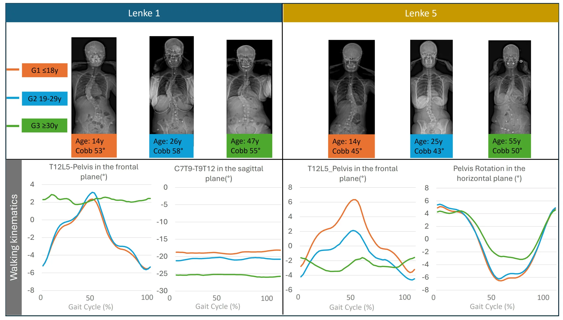
As submitted with the abstract. Tap a figure to open it at full size.