
Growing Spine
Evaluation of MRI “CT-like” Imaging for Pedicle Diameter Measurement in Adolescent Idiopathic Scoliosis
- The Jikei University School of Medicine, Tokyo, Japan
Abstract
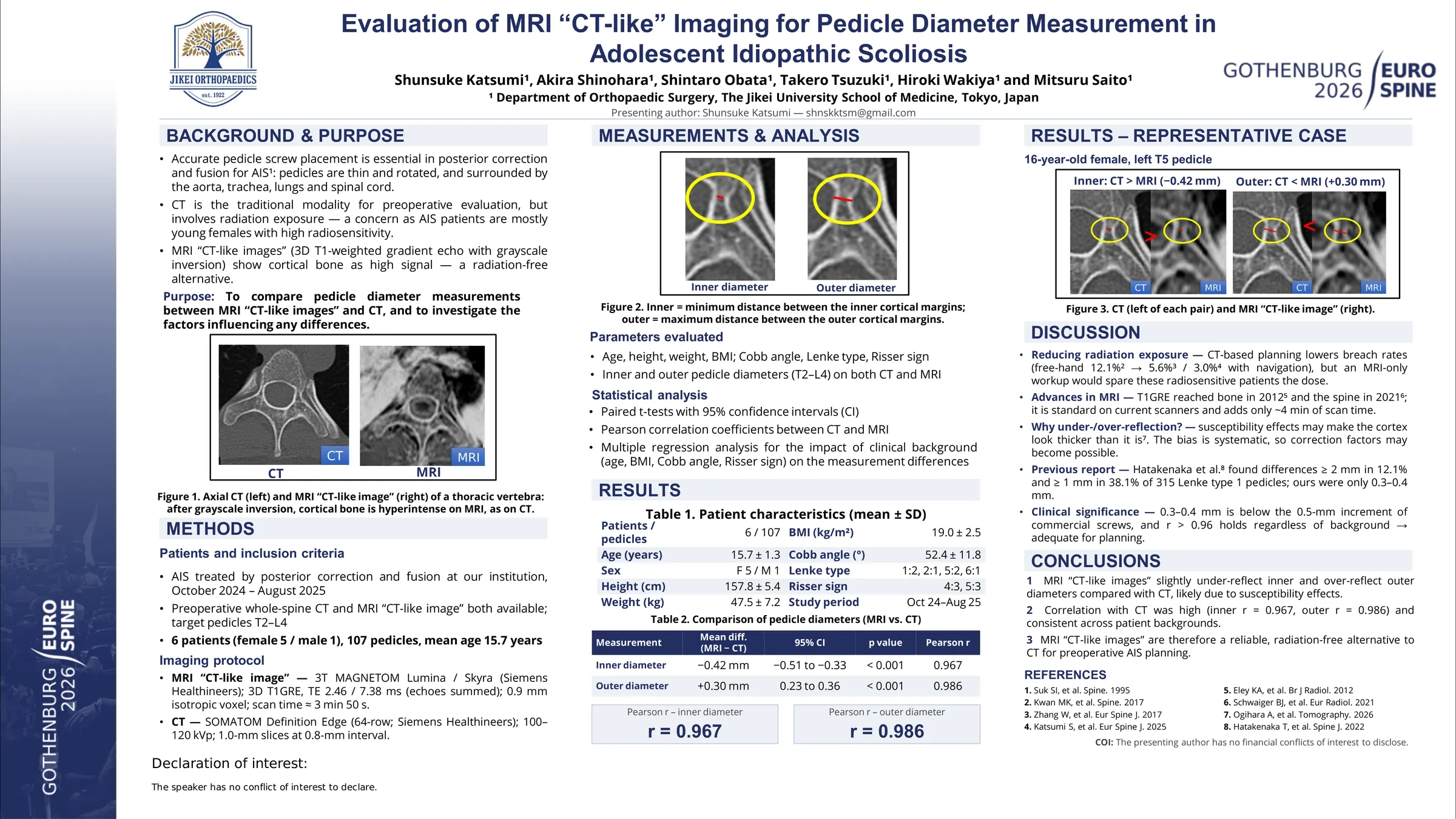
Accurate pedicle screw placement is essential in posterior spinal fusion for adolescent idiopathic scoliosis (AIS), as pedicle morphology directly influences screw size selection and trajectory planning. Preoperative computed tomography (CT) is widely used for detailed pedicle assessment; however, radiation exposure is a particular concern in adolescents, who may undergo repeated imaging during diagnosis and follow-up. Recently, MRI-based reconstruction techniques generating “CT-like images” have been introduced as a radiation-free alternative capable of visualizing bony structures with high resolution. Although preliminary reports suggest promising agreement with CT, quantitative validation in AIS pedicle assessment remains limited. This study aimed to compare inner and outer pedicle diameter measurements between MRI CT-like images and conventional CT and to investigate whether clinical factors influence any measurement discrepancies.
We retrospectively analyzed 107 pedicles (T2–L4) from 6 AIS patients (mean age 15.7 years) who underwent posterior spinal fusion between October 2024 and August 2025. All patients underwent both preoperative CT and MRI with CT-like image reconstruction. Inner and outer pedicle diameters were measured at corresponding vertebral levels using standardized axial planes.Paired t-tests were used to compare measurements between modalities, and 95% confidence intervals (CI) were calculated. Agreement was assessed using Pearson correlation coefficients. Multiple regression analysis evaluated the potential influence of age, body mass index (BMI), Cobb angle, and Risser sign on measurement differences.
MRI CT-like images demonstrated significantly smaller inner pedicle diameters compared with CT (mean difference −0.42 mm; 95% CI −0.51 to −0.33; p<0.001) and significantly larger outer diameters (mean difference +0.30 mm; 95% CI 0.23 to 0.36; p<0.001).Despite these systematic differences, strong positive correlations were observed for both inner (r=0.967) and outer diameters (r=0.986), indicating high linear agreement. Multiple regression analysis revealed no significant associations between measurement discrepancies and patient background factors.
Although MRI CT-like images slightly underestimate inner pedicle diameter and overestimate outer diameter compared with CT, the strong correlation and consistent measurement pattern across clinical backgrounds suggest reliable morphological assessment. Considering the importance of minimizing radiation exposure in adolescents with AIS, MRI CT-like imaging may represent a clinically feasible radiation-free alternative for preoperative pedicle evaluation.