
Growing Spine
Evaluating CT-Like MRI as a Radiation-Free Alternative for Preoperative Planning in Spinal Deformity Surgery
- KU Leuven, Leuven, Belgium
- UZ Leuven, Leuven, Belgium
Abstract
Computed tomography remains the reference standard for detailed osseous visualization in spinal deformity surgery. However, repeated CT imaging exposes patients particularly adolescents with idiopathic scoliosis (AIS) to cumulative ionizing radiation. While synthetic CT approaches derived from MRI have been proposed, many rely on proprietary AI-based tools that introduce financial, regulatory, and logistical barriers. An alternative strategy is CT-like MRI, which enhances bone contrast using conventional MRI acquisitions combined with straightforward post-processing, enabling in-house generation without specialized software dependencies. This study assessed whether CT-like MRI can achieve comparable accuracy to CT for surgical planning and geometric evaluation.
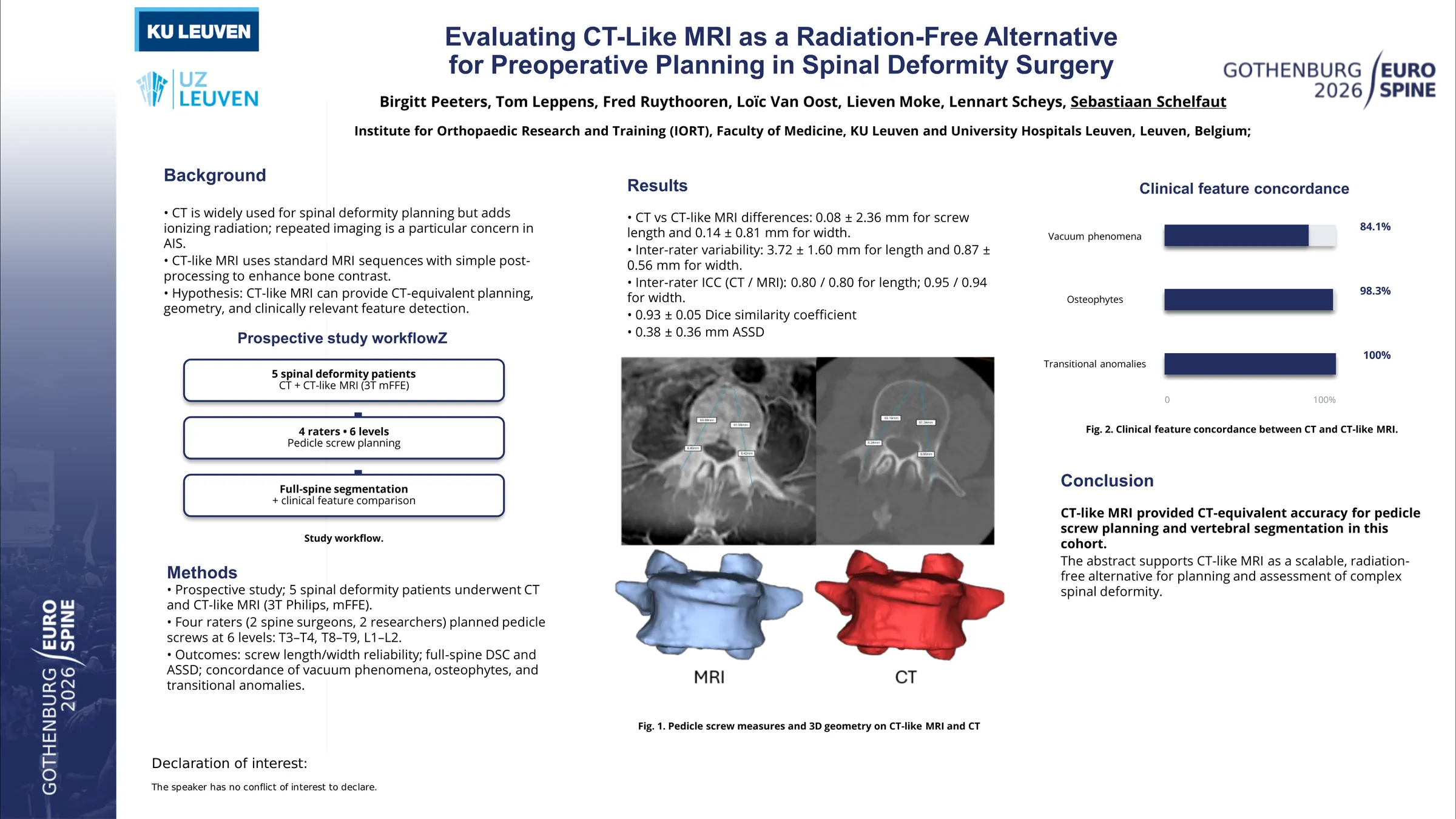
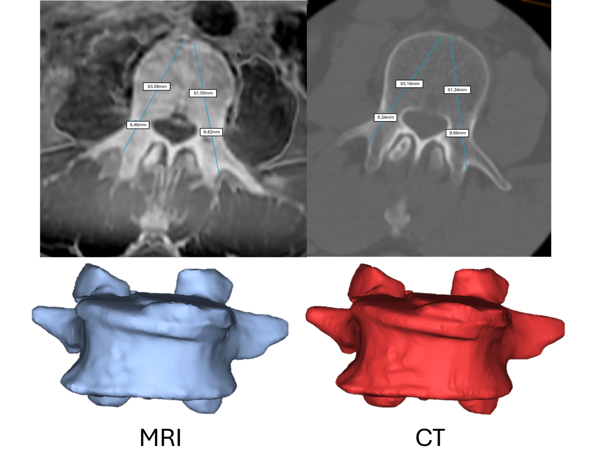
Five patients with spinal deformities underwent both conventional CT and CT-like MRI (3T Philips, mFFE sequence). Four independent raters (two spine surgeons, two researchers) performed pedicle screw planning at six predefined vertebral levels (T3–T4, T8–T9, L1–L2), measuring optimal screw length and diameter on both imaging modalities.
Agreement between modalities and raters was quantified using mean differences and intraclass correlation coefficients (ICC). Full-spine vertebral segmentation was evaluated to determine geometric consistency using the Dice similarity coefficient (DSC) and average symmetric surface distance (ASSD). Clinically relevant osseous features including vacuum phenomena, osteophytes, and transitional vertebral anomalies were assessed for inter-modality concordance.
Differences between CT and CT-like MRI were minimal: 0.08 ± 2.36 mm for screw length and 0.14 ± 0.81 mm for width. These discrepancies were smaller than observed inter-rater variability (3.72 ± 1.60 mm for length; 0.87 ± 0.56 mm for width). Inter-rater reliability was comparable across modalities (CT/MRI ICC: 0.80/0.80 for length; 0.95/0.94 for width).
Segmentation accuracy demonstrated high geometric fidelity (DSC 0.93 ± 0.05; ASSD 0.38 ± 0.36 mm). Detection agreement for clinical features was strong, with concordance rates of 84.1% for vacuum phenomena, 98.3% for osteophytes, and 100% for transitional anomalies.
CT-like MRI demonstrated equivalent performance to CT for pedicle screw planning, vertebral geometry assessment, and detection of clinically relevant osseous features. Given the increasing reliance on precision planning, robotic instrumentation, and patient-specific modeling in spinal deformity surgery, CT-like MRI represents a feasible, radiation-free imaging alternative that can be implemented without proprietary AI infrastructure.
Figures and tables
As submitted with the abstract. Tap a figure to open it at full size.