
Cervical & Neural Pathologies
Dynamic MRI in Cervical Myelopathy: Factors Associated with New Compression Findings
- Hospital Italiano de Buenos Aires, Buenos Aires, Argentina
Abstract
Cervical spondylotic myelopathy is the compression of the spinal cord secondary to degenerative narrowing of the canal. Dynamic cervical magnetic resonance imaging (MRI) can reveal new compressions that are not evident in neutral cervical MRI. This study aimed to identify radiographic and MRI factors associated with increased spinal cord compression when comparing neutral and dynamic MRI.
This was a retrospective comparative cohort study from 2017 to 2024, encompassing all patients with symptoms of spondylotic cervical myelopathy who underwent both dynamic and neutral cervical MRI and cervical X-rays.
We collected variables from neutral and dynamic lateral cervical X-rays. Neutral MRI variables included Goutallier classification, the canal diameter at C7, and the spinal cord-to-canal ratio (CCR%) measured on axial and sagittal MRI, respectively. Additionally, we recorded disc degeneration according to the Pfirrmann classification, intramedullary T2 high-intensity signal (HIS), facet joint degeneration, and the presence of facet effusion. Anterior and posterior spinal cord compression were graded using the Muhle scale.
A cervical compression change was defined as an increase of 2 grades or more in the Mulhe classification from neutral to dynamic on MRI.
We calculated the proportion of cervical compression changes between neutral and dynamic cervical MRI. Subsequently, we analyzed the risk of cervical compression change and compared each cervical level by clustering them into groups with and without compression changes. We performed mixed-effects logistic regression analyses with random intercepts for patients to account for multiple cervical levels per subject. Odds ratios with 95% confidence intervals were calculated.
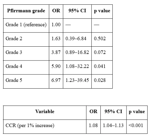
82 patients were included, and 57 (69.5%) had a cervical compression change at some level. When comparing cervical levels, Pfirrmann grades 4 and 5 were significantly associated with compression changes, with ORs of 5.90 (95% CI: 1.08–32.22; p=0.041) and 6.97 (95% CI: 1.23–39.45; p=0.028), respectively. The CCR also showed a strong association (OR=1.08; 95% CI: 1.04–1.13; p<0.01). Table 1.
Dynamic cervical MRI detected new levels of spinal cord compression in nearly 70% of cases. Higher Pfirrmann grades and increased CCR were significantly associated with these changes. These findings underscore the value of dynamic MRI in detecting hidden compression and guiding clinical decision-making in cervical myelopathy.
Figures and tables
As submitted with the abstract. Tap a figure to open it at full size.