
Trauma
Comparison of Simulation Navigation and Conventional AO-Method for Sacroiliac Screw Placement in Dorsal Pelvic Ring Fractures
- Kantonsspital Baselland, Bruderholz, Switzerland
- Kantonsspital Baselland Bruderholz, Bruderholz, Switzerland
Abstract
Safe sacroiliac (SI) screw placement in dorsal pelvic ring fractures, particularly in elderly patients with fragility fractures and sacral dysmorphism, remains technically demanding under conventional fluoroscopic guidance. This study aimed to compare the efficacy and accuracy of a CT-based three-dimensional (3D) simulation navigation method with the conventional AO fluoroscopy-based technique. The primary focus was to evaluate differences in operative time, screw placement accuracy, and overall surgical outcomes, with the goal of improving precision and patient safety in SI screw fixation.
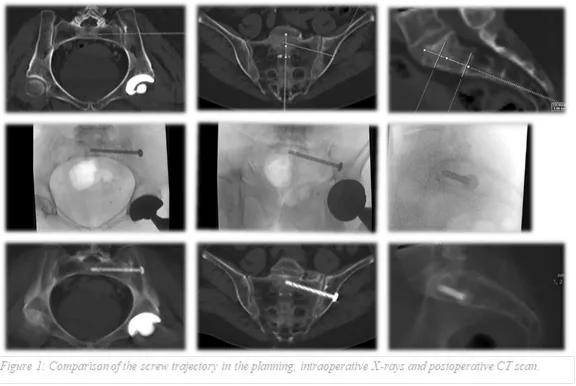
A total of 39 patients requiring dorsal pelvic ring stabilization with a dysmorphic sacrum using screw osteosynthesis were included in this study. Fourteen patients (25 screws) were treated using the conventional AO fluoroscopy-based technique, while 25 patients (41 screws) underwent screw placement using a CT-based simulation navigation method. In the simulation navigation group, preoperative planning included computed tomography with multiplanar reformation to identify anatomical landmarks and define safe intraosseous screw trajectories. In the conventional group, screw placement was performed under standard fluoroscopic guidance without preoperative trajectory simulation. Intraoperatively, the navigation group followed the preoperatively simulated screw pathway, whereas the conventional group relied solely on conventional fluoroscopic imaging.
The mean operative time was 84 minutes per patient (42 minutes per screw) in the conventional AO group compared with 58 minutes per patient (33 minutes per screw) in the navigation simulation group, representing a reduction of 26 minutes per patient and 9 minutes per screw. Partial screw malposition occurred in 2 screws (8%) in the AO group and in 1 screw (2.4%) in the navigation group. Postoperative CT analysis demonstrated improved screw alignment in the navigation group, with minimal deviation from the planned trajectory (mean angular deviation: 2.6° in the inlet view, 4.4° in the outlet view, and 9.7° in the sagittal plane). No neurologic complications were observed in either group.
The simulation navigation method significantly reduced operative time and maintained superior accuracy compared to the conventional AO-method for dorsal pelvic ring stabilization. With no neurologic defficiencies in either group, the navigation simulation method demonstrated enhanced efficiency in screw placement. This approach, utilizing preoperative planning and intraoperative guidance, appears to be a promising alternative for optimizing surgical outcomes in dorsal pelvic ring fractures in hospitals without expensive navigation systems.
Figures and tables
As submitted with the abstract. Tap a figure to open it at full size.