
Basic Science & Economics
Barycentrometric analysis of sagittal flexion moments in ASD: role of ODHA in identifying abnormal mechanical loading
- Saint Joseph University of Beirut, Beirut, Lebanon
- Arts et Métiers, Paris, France
Abstract
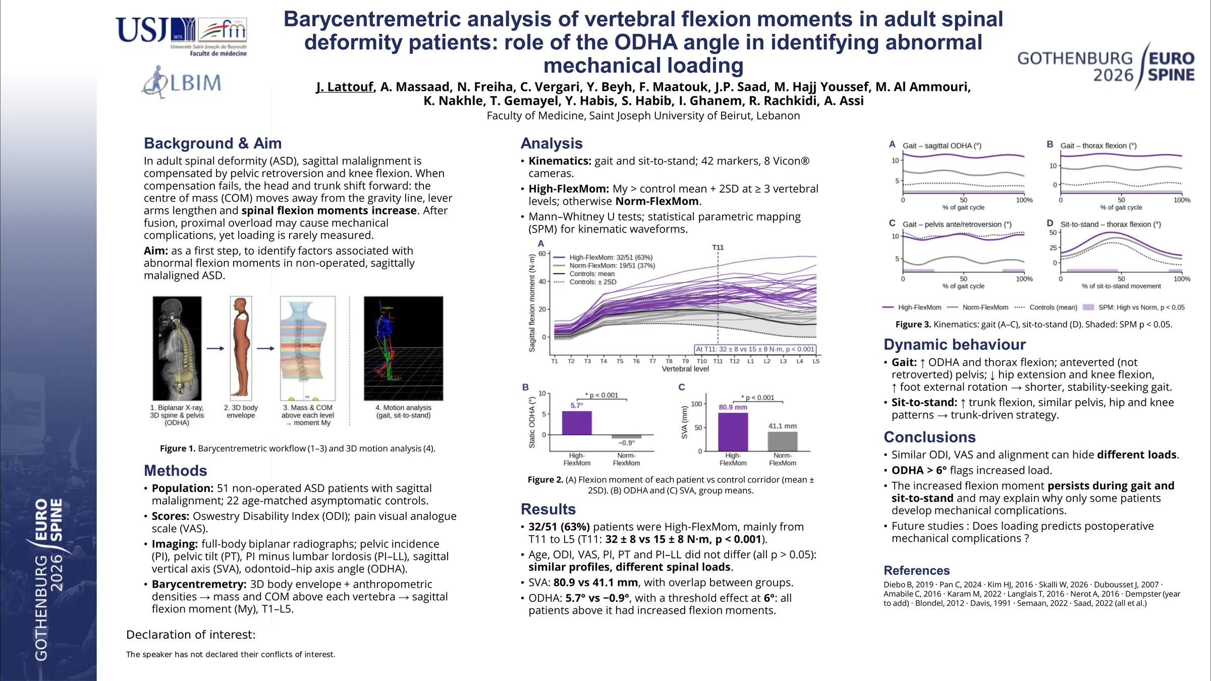
Postoperative mechanical complications in ASD occur in over 40% of cases and remain poorly understood through conventional radiographic analysis alone. These complications seem to arise from mechanical overload of the spinal segments proximal to the instrumentation, resulting in rod fracture or screw loosening. Quantifying spinal loads, particularly flexion moments, is essential to better understand these mechanisms. Biplanar imaging combined with 3D reconstruction and barycentrometric analysis allows quantification of vertebral moments at each level, providing an objective evaluation of sagittal loading. The aim was to quantify flexion moments at vertebral levels in ASD patients with sagittal malalignment and to identify the determinants associated with abnormal moment patterns.
51 ASD patients with sagittal malalignment (PT>25° and/or PI–LL>10° and/or SVA>50mm) filled QOL questionnaires (ODI and VAS for pain). All patients underwent standing full-body biplanar radiographs followed by 3D reconstruction of the spine, pelvis, and external envelope, to calculate spinopelvic and barycentrometric parameters. Using previously published and validated techniques, moment components of the upper segments at each vertebral level were calculated along the three axes. Patients were classified as High-FlexMom if their flexion moment exceeded normative values (mean+2SD of 90 asymptomatic controls) at least at 3 vertebral levels; otherwise, they were classified as Norm-FlexMom. Spinopelvic parameters and QOL scores were compared between groups.
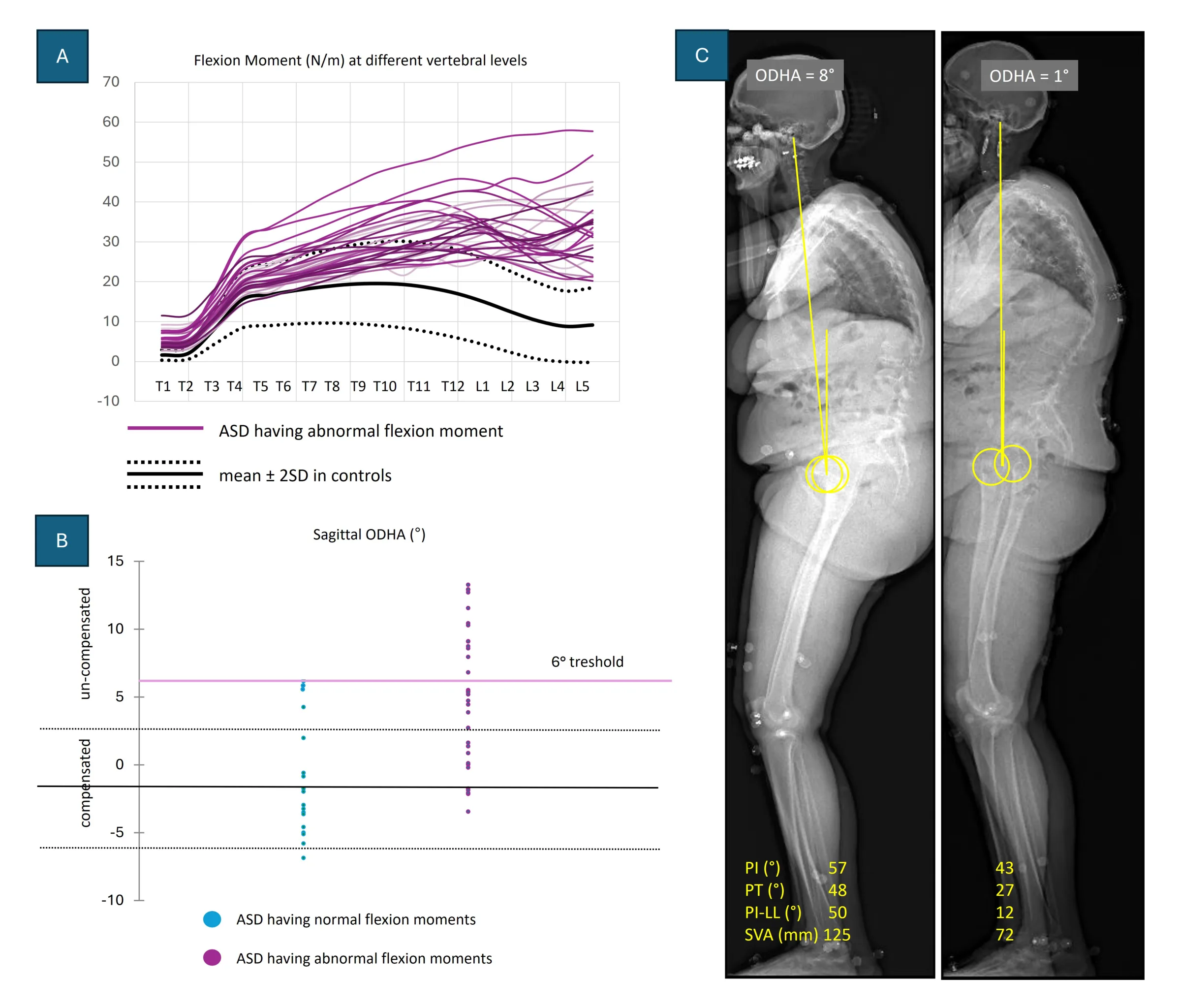
32/51 patients were classified as High-FlexMom, showing increased flexion moments from T11 to L5 (30 ±8N/m; Fig1.A). 19/51 patients were classified as Norm-FlexMom. Age (65y), ODI (37), VAS for pain (7) and spinopelvic parameters were similar between groups, including PI (56°), PT (30°), and PI–LL (18°; all p>0.05). Patients in the High-FlexMom group had greater SVA (81 vs 41mm, p=0.005) and sagittal ODHA (5.5 vs 0°, p<0.001) compared with the Norm-FlexMom group. While SVA values overlapped between groups, ODHA showed a clear threshold effect at 6° (Fig.1.B), above which all patients had abnormally increased flexion moments.
Surprisingly, ASD patients with increased sagittal flexion moments showed similar QOL scores and spinopelvic parameters compared to patients presenting normal flexion moments. The main difference was a greater forward inclination. A sagittal ODHA threshold of 6° distinguished abnormal flexion moment patterns (example in Fig1.C). Barycentrometric analysis may therefore provide clinically relevant mechanical information that is not captured by conventional alignment parameters alone. This approach could help identify patients with abnormal sagittal loading and may contribute to a better understanding of mechanical factors potentially involved in postoperative mechanical complications.
Figures and tables
As submitted with the abstract. Tap a figure to open it at full size.