
Degenerative Thoracolumbar
Axial loaded MRI in surgically treated LSS patients, a report from the NORDSTEN study
- Akershus University Hospital, Lorenskog, Norway
- Møre and Romsdal Hospital Trust, Ålesund, Norway
- Institute of Clinical Sciences, Sahlgrenska Academy, University of Gothenburg, Gothenburg, Sweden
- University of Gothenburg, Gothenburg, Sweden
Abstract
Standard lumbar MRI may fail to capture how weight-bearing affects the narrowing of the spinal canal and neural foramina. While the diagnostic value of axial loading is documented in primary lumbar spinal stenosis (LSS), its value after decompression is unclear. This study evaluates the utility of axial-loaded MRI (AL-MRI) in assessing central and foraminal stenosis in LSS patients five years post-decompression.
This study included patients from the NORwegian Degenerative spondylolisthesis and Spinal STENosis (NORDSTEN-SST) study, five years after undergoing one of three minimally invasive decompression techniques for LSS: unilateral laminotomy with cross-over, bilateral laminotomy, or spinous process osteotomy. Imaging was performed using supine MRI both with and without axial loading (AL-MRI). Two radiologists independently assessed the dural sac cross-sectional area (DSCA), Schizas and Lee grading (L2-L5), and an interobserver reliability test was performed. The association between loading‑induced DCSA changes and patient‑reported outcome measures (PROMs), including the Oswestry Disability Index (ODI) and Numeric Rating Scale, was analysed at five years post‑surgery.
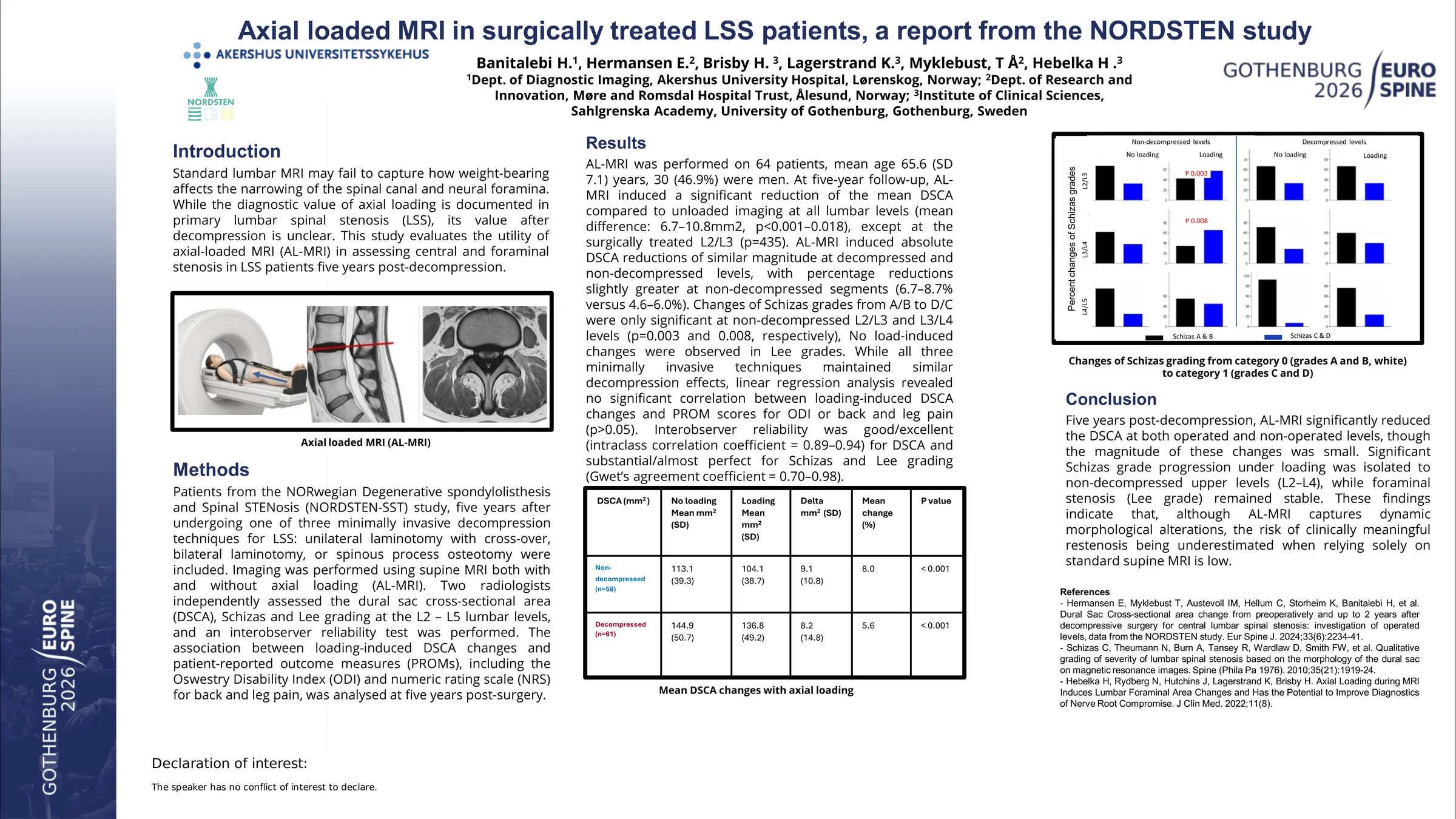
In total, 64 patients underwent AL-MRI. Mean age was 65.6 (SD 7.1) years and 30 (46.9%) were men. At five-year follow-up, AL-MRI induced a significant reduction of the mean DSCA compared to unloaded imaging at all lumbar levels (mean difference: 6.7–10.8mm2, p<0.001–0.018), except at the surgically treated L2/L3 (p=435). AL-MRI induced absolute DSCA reductions of similar magnitude at decompressed and non‑decompressed levels, with percentage reductions slightly greater at non‑decompressed segments (6.7–8.7% versus 4.6–6.0%). Changes of Schizas grades from A/B to D/C were only significant at non-decompressed L2/L3 and L3/L4 levels (p=0.003 and 0.008, respectively), while no load‑induced changes were observed in Lee grades. While all three minimally invasive techniques maintained similar decompression effects, linear regression analysis revealed no significant correlation between loading-induced DSCA changes and PROM scores for ODI or back and leg pain (p>0.05). Interobserver reliability was good/excellent (intraclass correlation coefficient = 0.89–0.94) for DSCA and substantial/almost perfect for Schizas and Lee gradings (Gwet’s agreement coefficient = 0.70–0.98).
Five years post-decompression, AL-MRI significantly reduced the DSCA at both operated and non-operated levels, though the magnitude of these changes was small. Significant Schizas grade progression under loading was isolated to non-decompressed upper levels (L2–L4), while foraminal stenosis (Lee grade) remained stable. These findings indicate that, although AL-MRI captures dynamic morphological alterations, the risk of clinically meaningful restenosis being underestimated when relying solely on standard supine MRI appears low.