
Basic Science & Economics
Are hyperhydration and endplate-anulus integrity potential key drivers of cervical disc herniation? – An ex vivo study
- Ulm University Medical Centre, Ulm, Germany
- University of Health Sciences, Bochum, Bochum, Germany
- Charité – University Medicine Berlin, Berlin, Germany
- German Sport University, Cologne, Germany
- Sahlgrenska University Hospital, Gothenburg, Sweden, Sweden
- University of Birmingham, Birmingham, UK, United Kingdom
- Johnson Space Center, Houston, United States of America
- Aeromedical Centre, Helsinki, Finland
Abstract
Pathomechanisms underlying cervical intervertebral disc (IVD) herniation remain poorly understood, including the role of structural tissue damage to herniation susceptibility. While complex loading promotes herniation in lumbar IVDs [1], its relevance to the cervical spine is unclear. Astronauts show a 21-fold higher risk of cervical IVD herniation, potentially linked to microgravity-induced IVD hyperhydration [2]. Building on these considerations, the aim of this study was to assess whether swelling-induced changes in intradiscal pressure (IDP), range of motion (ROM), and neutral zone (NZ), in combination with endplate–anulus compromise, influence susceptibility to cervical IVD herniation under complex loading.
Six fresh-frozen human cervical motion segments (1x C3-C4, 3x C5-C6, 2x C6-C7; 17–42 years; 2 female / 4 male) were hydrated in distilled water (to obtain hyperhydration) for 14h, while IDP was monitored using fibreoptic pressure transducers. A paramedial posterior defect at the cranial endplate-anulus transition was created under radiographic control. Specimens were then subjected to 3000 cycles of complex loading at 1 Hz in a dynamic disc-loading simulator [3], consisting of 100 N axial compression combined with flexion/extension (FE), lateral bending (LB), and axial rotation (AR) within physiological ROM [4]. Flexibility was evaluated before and after hydration and after complex loading using pure moments of 2.5 Nm in a spine tester. Friedman test with Bonferroni correction was performed in SPSS29.
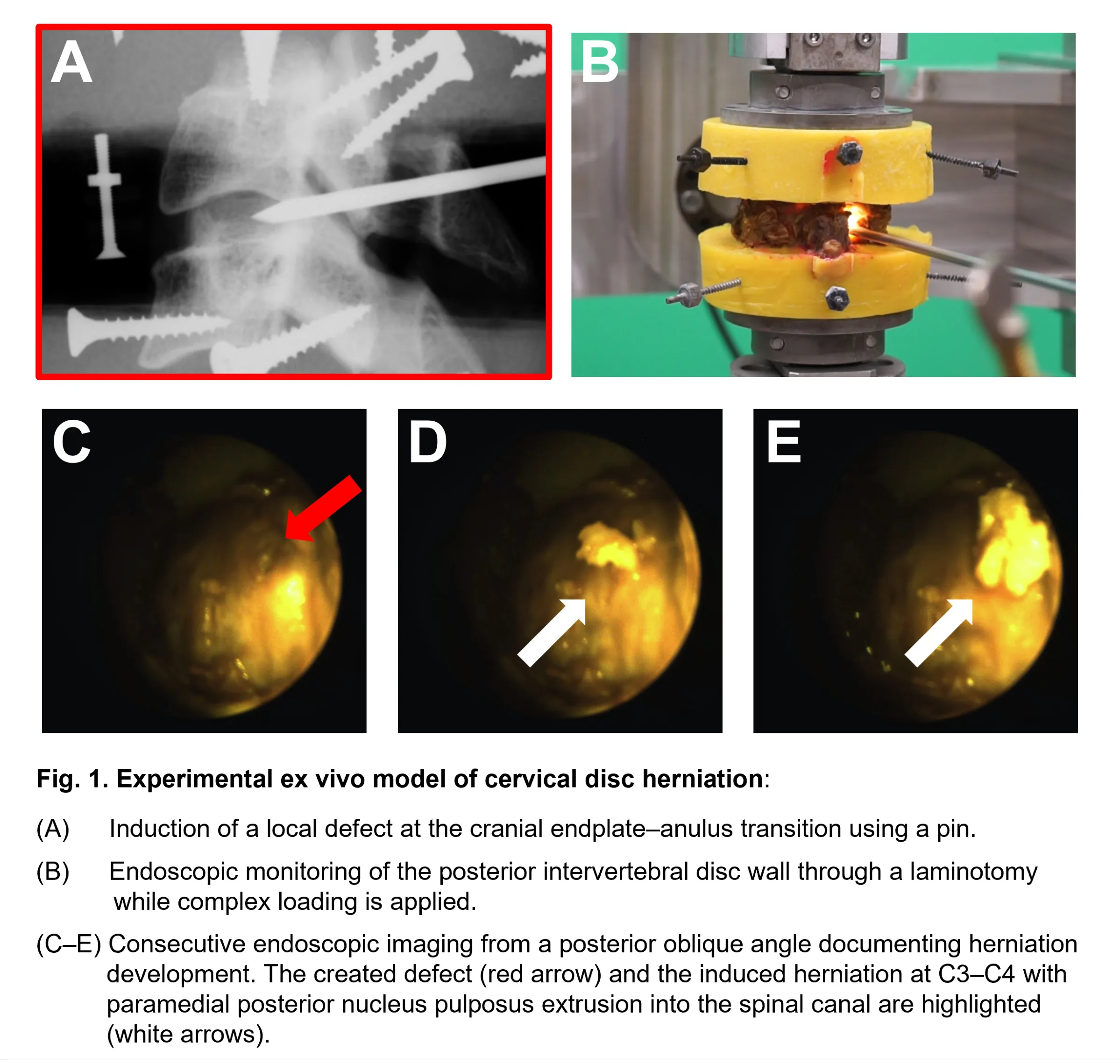
Hyperhydration increased median intrinsic IDP by +243% (p<0.05) and reduced median NZ (FE: -46%, LB: -56%; p<0.05). Complex loading induced herniation in one of six IVDs, characterized by posterior, paramedial extrusion of nucleus pulposus material into the spinal canal at the cranial endplate defect site (Fig. 1). After complex loading, both median ROM (FE: +36%, LB: +56%, AR: +31%) and median NZ (FE: +148%, LB: +216%, AR: +138%) increased significantly compared to the hyperhydrated condition (p < 0.05).
Hyperhydration increases IDP and reduces NZ, indicating elevated stiffness and mechanical stress within IVD tissues. As IDP reflects the load acting on the IVD, this hydration-related pressure increase together with complex loading may therefore promote cervical IVD herniation. Conversely, a compromise at the endplate–anulus interface did not consistently induce herniation, suggesting that this structural disruption alone is unlikely to be a decisive factor. Overall, the results show that hyperhydration increases IDP and IVD stiffness, making the cervical IVD more susceptible to herniation during high‑risk movements.
Acknowledgements: Funded by the German Research Foundation (DFG, WI1352/14-4) and the German Aerospace Centre (DLR, 50WB2424D). References: [1] Slater et al. (2025). Eur Spine J 34, 4353-68; [2] Belavy et al. (2016). Eur Spine J 25,144-154; [3] Wilke et al. (2016). Eur Spine J 25, 1363-1372; [4] Anderst et al. (2015). J Biomech 48, 1286-93.
Figures and tables
As submitted with the abstract. Tap a figure to open it at full size.