
Complications & Epidemiology
Anterior-to-Psoas (ATP) Lumbar Interbody Fusion at L4-L5: Radiographic Outcomes and Influence of Psoas Morphology
- St. Joseph's University Medical Center, Paterson, United States of America
- St. Joseph's University Medical Center, Wayne, United States of America
Abstract
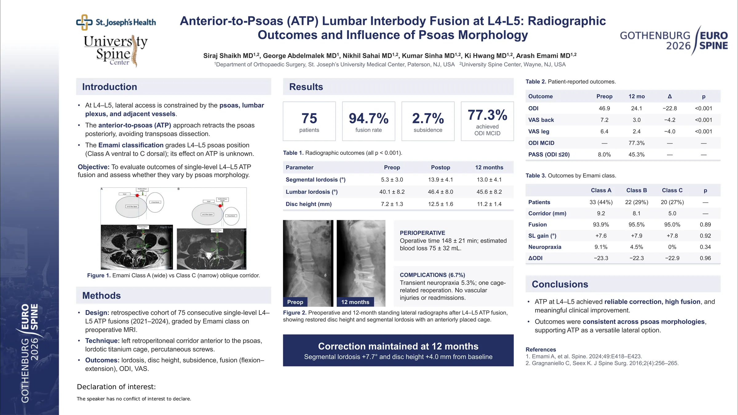
The anterior-to-psoas (ATP) approach provides a unique corridor to the lumbar spine, allowing direct disc access without psoas violation and preservation of posterior elements. This enables ATP to leverage advantages of both lateral and oblique lumbar interbody fusion techniques while potentially mitigating lumbar plexus and vascular injury. Despite increasing adoption, limited data exist evaluating ATP as an independent technique, and optimal patient selection criteria remain unclear. A novel psoas morphology classification system has been introduced to guide lateral access strategies; however, its relationship to ATP outcomes has not been established. The purpose of this study was to evaluate radiographic, perioperative, and patient-reported outcomes following single-level L4–L5 ATP fusion and to assess whether complication patterns vary according to preoperative psoas morphology.
A retrospective cohort study was performed of 48 adult patients undergoing single-level L4–L5 ATP fusion between January 2021 and December 2024. Preoperative MRI was used to classify psoas morphology (Class A, B, or C). Standing radiographs and dynamic flexion–extension imaging were reviewed postoperatively and at latest follow-up to assess alignment, disc height, subsidence, and fusion status.
Psoas morphology distribution was Class A (43.8%), Class B (29.2%), and Class C (27.1%), with corresponding mean oblique corridor widths of 9.3 ± 1.4 mm, 8.0 ± 1.2 mm, and 4.9 ± 1.1 mm, respectively. Radiographic fusion was achieved in 91.7% of patients, with subsidence observed in 2 cases (4.2%). Segmental lordosis improved from 5.2° preoperatively to 12.9° at one year, lumbar lordosis from 39.8° to 45.5°, and disc height from 7.1 mm to 11.1 mm, with minimal loss over time. One patient experienced hardware failure requiring reoperation. No readmissions, infections, or vascular injuries occurred. Transient neuropraxia was observed in three patients, most commonly in Class A morphology, though differences were not statistically significant. Mean ODI improved from 46.8 ± 13.2 preoperatively to 23.9 ± 13.5 at 12 months (p < 0.001), with 77.1% achieving MCID. Mean VAS back improved from 7.1 ± 1.6 to 2.9 ± 2.0 and VAS leg from 6.3 ± 2.0 to 2.3 ± 2.1 (both p < 0.001), with no significant differences across morphology classes.
Single-level L4–L5 ATP fusion demonstrates reliable radiographic correction, high fusion rates, meaningful improvement in patient-reported outcomes, and a low complication profile. Although underpowered for definitive subgroup comparisons, complication patterns aligned with anticipated anatomic risk profiles based on psoas morphology, while clinical improvement remained consistent across morphologic classes. These findings support the use of preoperative psoas morphology to guide lateral approach selection.