
Growing Spine
AI-Assisted Bone Age Estimation: Multicenter Validation and Comparison across Idiopathic Scoliosis Groups and Healthy Controls
- Afyonkarahisar Sağlık Bilimleri Üniversitesi (AFSÜ), Afyonkarahisar, Türkiye
- Ataşehir Florence Nightingale Hastanesi, Istanbul, Türkiye
- Nya Karolinska, Stockholm, Sweden
- Private, İzmir, Türkiye
- VMI Medical Park Hospital Kocaeli, İzmit, Türkiye
- Ondokuz Mayıs University, Samsun, Türkiye
- Adnan Menderes University, Aydın, Türkiye
- Anatolian Spine Study Group - ASSG, Istanbul, Türkiye
- Baltalimanı Kemik Hastalıkları Eğitim ve Araştırma Hastanesi, Levent Semt Polikliniği, Istanbul, Türkiye
- Istanbul Science University, Istanbul, Türkiye
Abstract
Accurate assessment of skeletal maturity is essential for predicting curve progression and optimizing treatment timing in idiopathic scoliosis (IS). Conventional bone age methods remain observer-dependent. Deep learning–based approaches offer automated and objective alternatives; however, validation across disease severity groups and populations is limited. This study aimed to validate an AI-assisted bone age estimation program in a Turkish multicenter cohort and to compare skeletal maturity patterns across IS severity groups and healthy controls.
A multicenter retrospective review was conducted across 8 centers in Turkey. The cohort included IS patients aged 8–18 years who underwent left hand–wrist radiography, along with age-matched healthy controls. Subjects were categorized as Group 1 (IS, Cobb 10–40°), Group 2 (IS, Cobb>40°, surgical candidates), and Group 3 (HC). Bone age was assessed using AI and by an experienced radiologist. Accuracy was evaluated using Pearson correlation, mean absolute error (MAE), and bias. Between-group comparisons were performed using ANOVA or Kruskal–Wallis tests, with subgroup analyses by sex and age (8–12, 13–15, and 16–18 years).
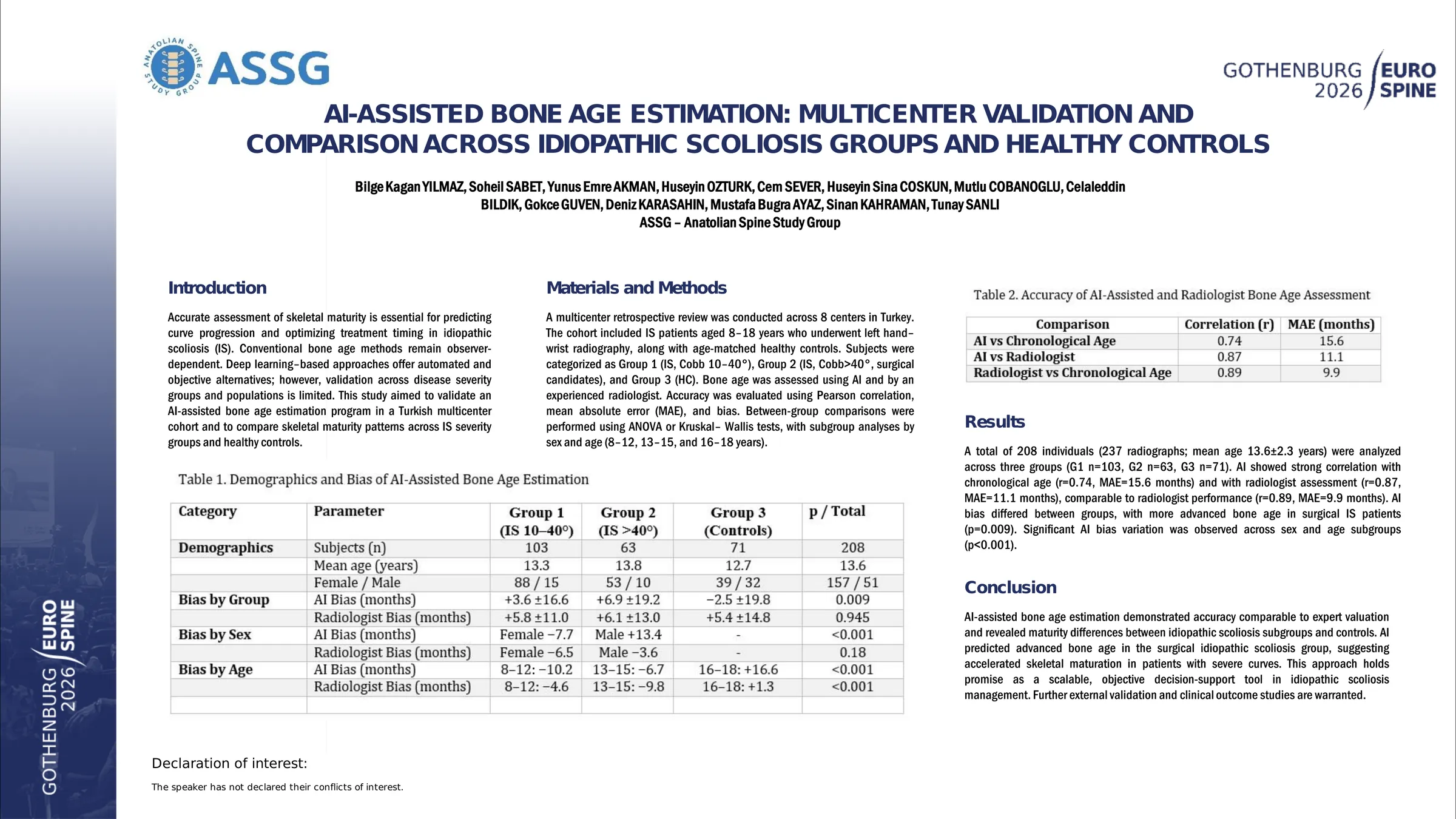
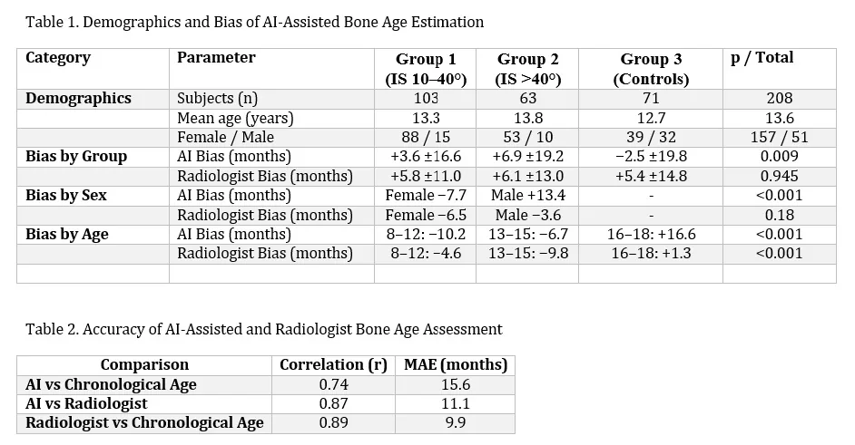
A total of 208 individuals (237 radiographs; mean age 13.6±2.3 years) were analyzed across three groups (G1 n=103, G2 n=63, G3 n=71). AI showed strong correlation with chronological age (r=0.74, MAE=15.6 months) and with radiologist assessment (r=0.87, MAE=11.1 months), comparable to radiologist performance (r=0.89, MAE=9.9 months). AI bias differed between groups, with more advanced bone age in surgical IS patients (p=0.009). Significant AI bias variation was observed across sex and age subgroups (p<0.001).
AI-assisted bone age estimation demonstrated accuracy comparable to expert valuation and revealed maturity differences between idiopathic scoliosis subgroups and controls. AI predicted advanced bone age in the surgical idiopathic scoliosis group, suggesting accelerated skeletal maturation in patients with severe curves. This approach holds promise as a scalable, objective decision-support tool in idiopathic scoliosis management. Further external validation and clinical outcome studies are warranted.
Figures and tables
As submitted with the abstract. Tap a figure to open it at full size.