
Degenerative Thoracolumbar
Voxel-wise MRI analysis reveals clinical and demographic associations in LSS
- Sahlgrenska University Hospital, Gothenburg, Sweden
- Møre and Romsdal Hospital Trust, Ålesund, Norway
- University of Gothenburg, Gothenburg, Sweden
- Akershus University Hospital, Lorenskog, Norway
- Sahlgrenska University Hospital, Gothenburg, Sweden, Sweden
- Haukeland University Hospital, Bergen, Norway
Abstract
Magnetic resonance imaging (MRI) is central to the evaluation of lumbar degenerative diseases such as lumbar spinal stenosis (LSS). However, radiological findings do not consistently reflect symptoms and many individuals with severe degeneration on MRI may report little or no symptoms. This image-symptom discrepancy represents an important research gap. Bridging this gap is essential for improving decision-making and tailoring treatment to individual patients.
This study aims to address this gap by using a holistic image and data-driven analysis approach, building upon methods such as Imiomics that have been proven successful in other research areas. Rather than focusing on predefined radiological metrics, complete MRI volumes from a large cohort of LSS patients were analysed with data-driven and voxel-wise methods to capture the effect of degeneration in the lumbar spine and neighbouring tissue.
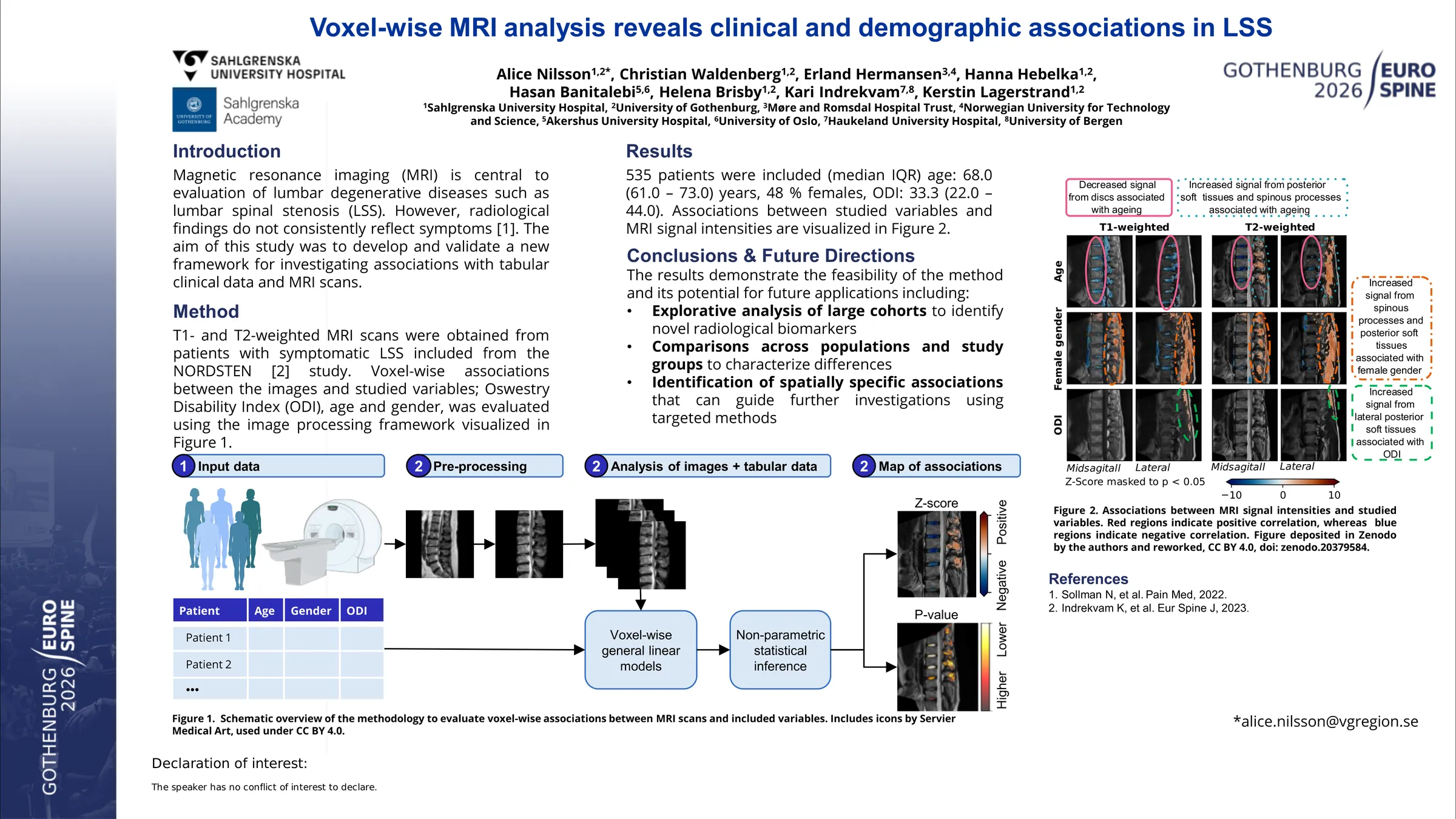
T1- and T2-weighted sagittal MRI scans were obtained from patients with symptomatic LSS scanned on 1.5 T MRI systems. T1- and T2-weighted images were processed separately and all sagittal slices from a single acquisition were treated as a single 3D volume. To enable direct voxel-wise comparison across patients and MRI examinations, volumes were pre-processed, including spine straightening, intersubject registration, and contrast normalization to a healthy reference scan.
General linear models were used to test the association between MRI voxel intensities and Oswestry Disability Index (ODI), age, and gender. Permutation testing (5 000 permutations) controlled the family-wise error rate at 5%. Significant associations were visualized using Z-score maps.
A total of 535 patients were included (median (IQR) age: 68.0 (61.0-73.0) years, ODI: 33.3 (22.0-44.0), and 48 % females), with a total of 526 T1-weighted and 508 T2-weighted volumes. No significant associations were found between MRI voxel-intensities and ODI, except for a localized T2 signal increase in the posterior spinal soft tissues. In contrast, both age and female gender were associated with MRI signal patterns. Higher age correlated with lower disc T1 and T2 signal intensities and increased T2 signal intensity in the posterior spinal soft tissue and spinous processes. The female gender was associated with increased T1 and T2 signal intensities in the posterior spinal soft tissues and spinous processes.
Voxel-wise MRI analysis showed only weak associations between ODI and MRI but demonstrated feasibility for detecting anatomical patterns and demographics in LSS that might be clinically relevant. This suggests that this method offers a valuable complementary tool to conventional radiological metrics for large-scale investigations of lumbar degenerative diseases.
Figures and tables
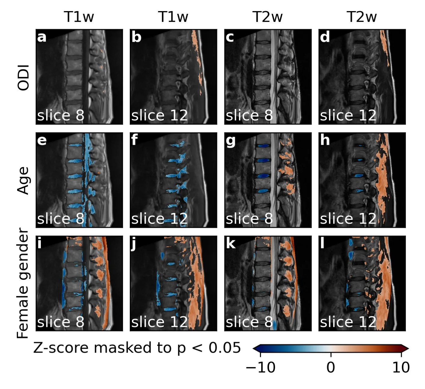
As submitted with the abstract. Tap a figure to open it at full size.