
Adult Deformity
Three Points Accurately Describe Sagittal Spinal Shape
- Vall d'Hebron University Hospital, Barcelona, Spain
- Dribia, Barcelona, Spain
- CHRU strasbourg - Les Hôpitaux Universitaires de Strasbourg , Strasbourg, France
- Vall d'Hebron Institut de Recerca (VHIR), Barcelona, Spain
- Acibadem University Maslak Hospital, Istanbul, Türkiye
- Chu De Bordeaux - Haut-Lévêque, Bordeaux, France
- Schulthess Klinik, Zurich, Switzerland
- La Paz University Hospital, Madrid, Spain
- University of Texas Health Science Center at San Antonio, Sant Antonio, United States of America
- Rady Children's Hospital, San Diego, United States of America
- . European Spine Study Group (Vall d'Hebron University Hospital, Barcelona, Spain
Abstract
Sagittal radiographs in individuals without disc degeneration or deformity suggest that the combination of pelvic incidence, L1PA, and T4PA can describe spinal shape and distribution of lordosis. However, the number of parameters to fully characterize sagittal shape is unclear. This study aimed to determine the minimum number of vertebral–pelvic angles (VPA) required to accurately describe global sagittal shape.
An AI-leveraged retrospective analysis was conducted using data from a prospective international multicenter database dedicated to ASD. Prospectively collected full-spine standing pre- and post-operative radiographs from 690 adults undergoing spinal deformity surgery were analyzed (81% female, average age 52 ± 19 years, preoperative Cobb 44.34º ± 24.04, GAP score 6.75 ± 4.42). A Machine-Learning automated vertebral centroid generation algorithm derived all centroids from C7 to S1 and the bicoxofemoral axis. VPAs from C7 to L5 were calculated.
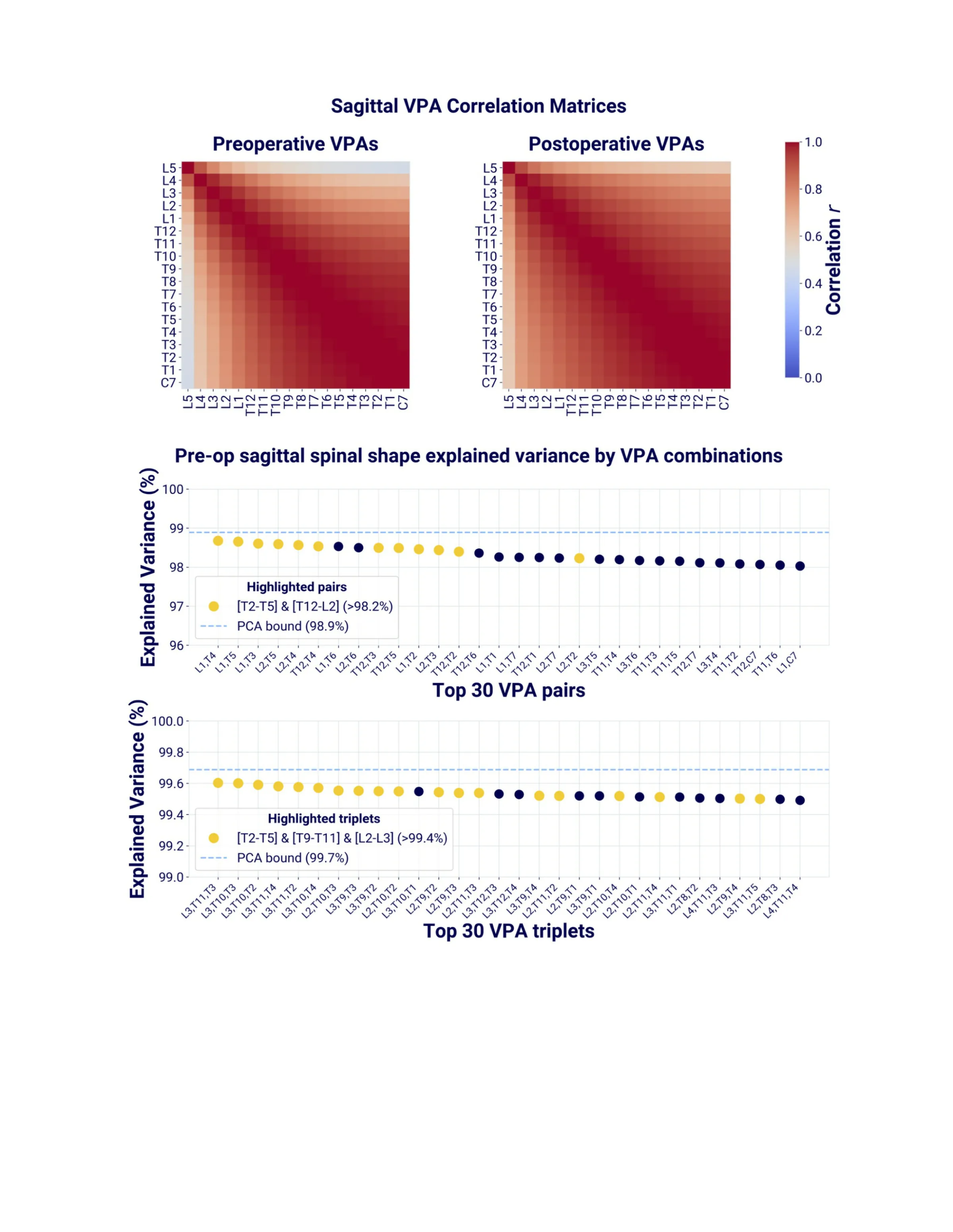
Linear correlations among VPAs were assessed. The proportion of sagittal spinal shape explained by subsets of VPAs was rigorously quantified via a robust metric to determine the minimum number required for accurate characterization, with principal component analysis (PCA) providing a reference for the maximum achievable explained variance.
Strong correlations were observed among all VPAs, with the highest correlations between adjacent vertebrae (r>0.89, p<0.001). The explained variance (R²) when using any VPA cranial to L1 for predicting the position of other VPAs was >85%. VPA pairs combining a VPA from the upper thoracic (T2-T5) and thoracolumbar (T12-L2) spine regions had the highest explained variance, accounting for >98% variability of the 16 (C7 to L5) remaining VPAs. Triplets combining upper thoracic (T2-T5) + lower thoracic (T9-T11) + mid lumbar (L2-L3) VPAs explain >99% of sagittal spinal shape. Estimates derived from preoperative and postoperative images were not significantly different.
Sagittal spinal shape can be accurately characterized using a small number of VPAs, with 2 VPAs capturing most of the variance. L1PA combined with the T4PA is among the most effective VPA pairs for describing overall spinal shape and lordosis distribution. These findings in ASD patients validate hypotheses previously generated in healthy controls, confirm vertebral pelvic angles and support radiomics-driven radiographic analysis for personalized surgical planning.
Figures and tables
As submitted with the abstract. Tap a figure to open it at full size.