
Degenerative Thoracolumbar
Segmental Lordosis Restoration at L5/S1 and Reciprocal Change in Short-Segment Fusion Using Oblique Lateral Interbody Fusion at L5–S1
- Kyoto University, Graduate School of Medicine, Kyoto, Japan
- Kyoto City Hospital, Kyoto, Japan
Abstract
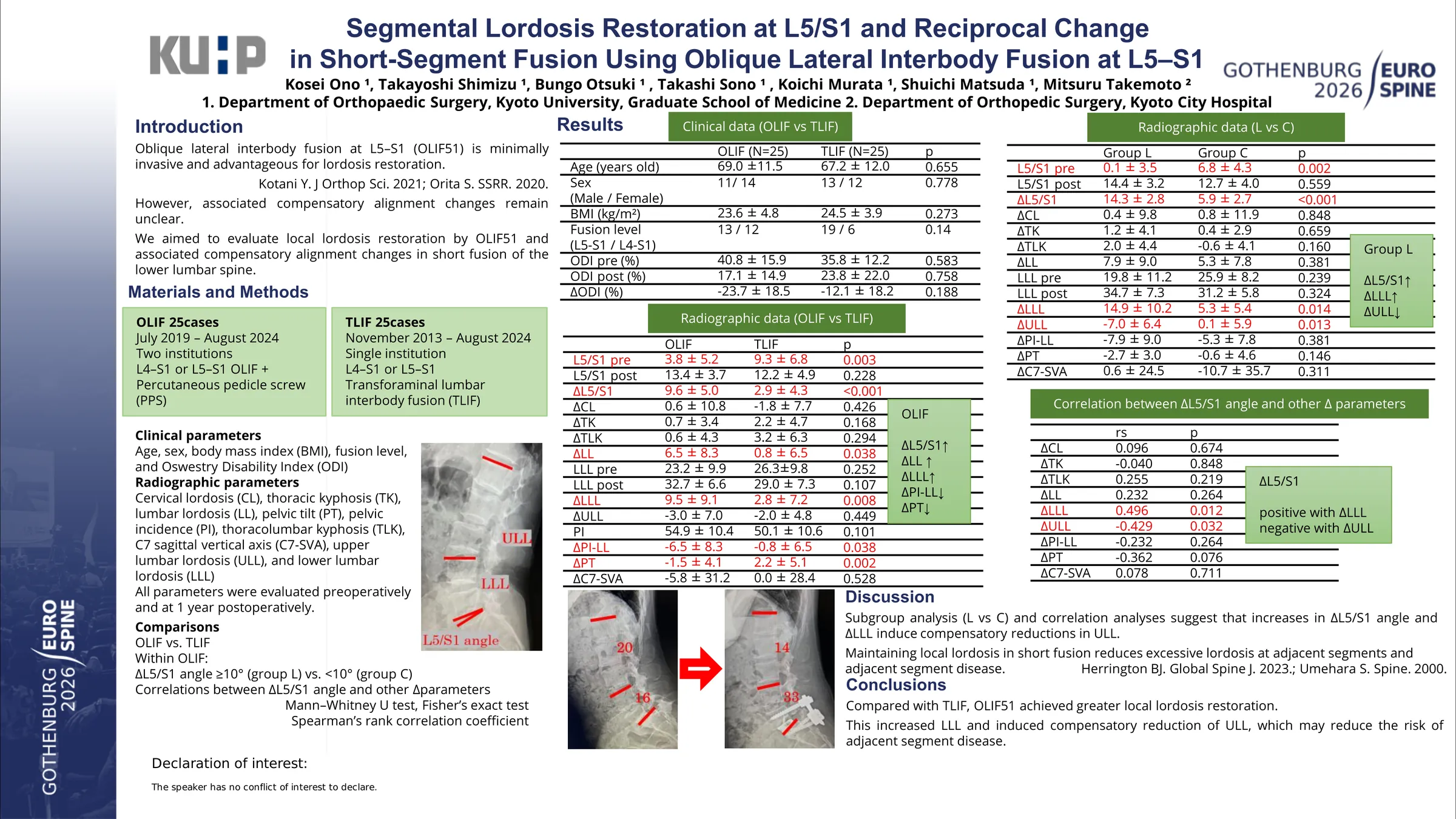
Restoration of segmental lordosis is important even in short-segment fusion, particularly at the lumbosacral junction, to optimize alignment and reduce mechanical stress on the remaining mobile segments. Oblique lateral interbody fusion at L5–S1 is a minimally invasive technique that can facilitate lordosis creation; however, its reciprocal impact on global spinal alignment remains unclear. This study evaluated L5–S1 segmental lordosis restoration and compensatory changes after short-segment fusion.
Patients who underwent short fusion at L5–S1 or L4–S1 with oblique lateral interbody fusion (O group, 2019–2024, two centers) or transforaminal lumbar interbody fusion (T group, 2013–2024, one center) were retrospectively reviewed. Inclusion criteria were no prior spinal fusion and at least 1-year follow-up. Twenty-five patients were analyzed in each group (O: 69.0±11.5 years, 14 women; T: 67.2±12.0 years, 12 women). Radiographic parameters and the Oswestry Disability Index (ODI) were compared. Radiographic measurements included L5–S1 segmental lordosis angle (SDA), cervical lordosis (CL), thoracic kyphosis (TK), thoracolumbar kyphosis (TLK), lumbar lordosis (LL), lower lumbar LL (LLL), upper LL (ULL), pelvic incidence (PI), PI–LL, pelvic tilt (PT), and C7 sagittal vertical axis (SVA), assessed preoperatively and at 1 year postoperatively. Subgroup analysis was performed in the O group by defining the lordosis-restored group (L group, change in L5-S1 SDA ≥10°, n=11) and the control group (C group, <10°, n=14). Correlations between change in L5-S1 SDA and other parameter changes were assessed.
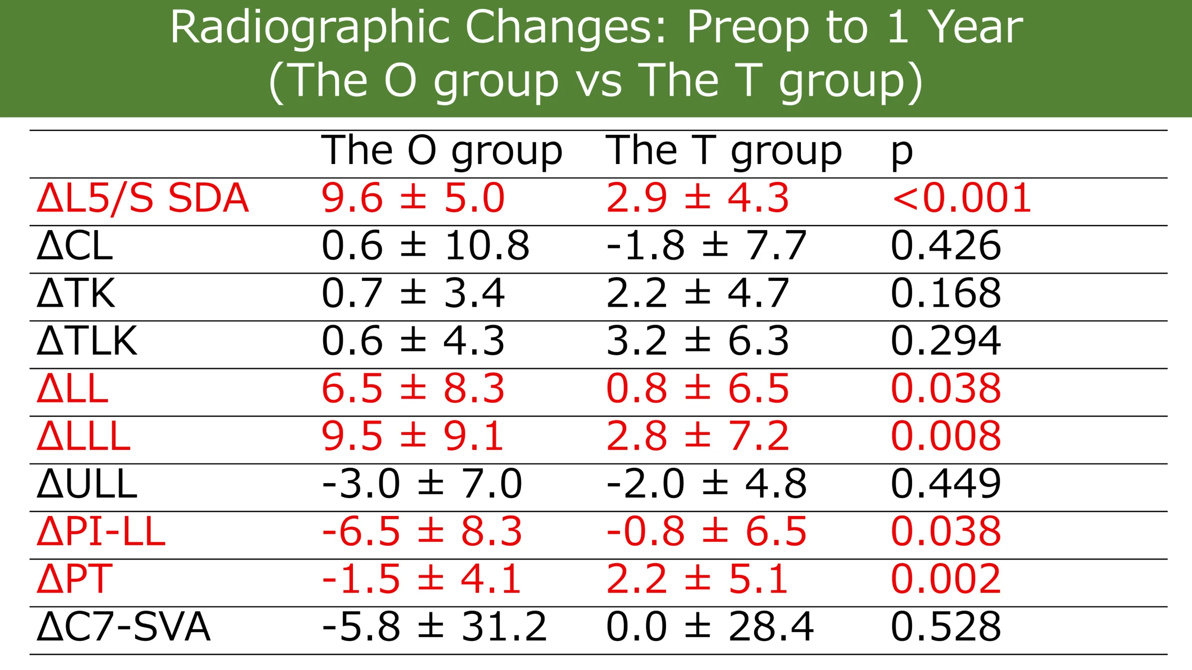
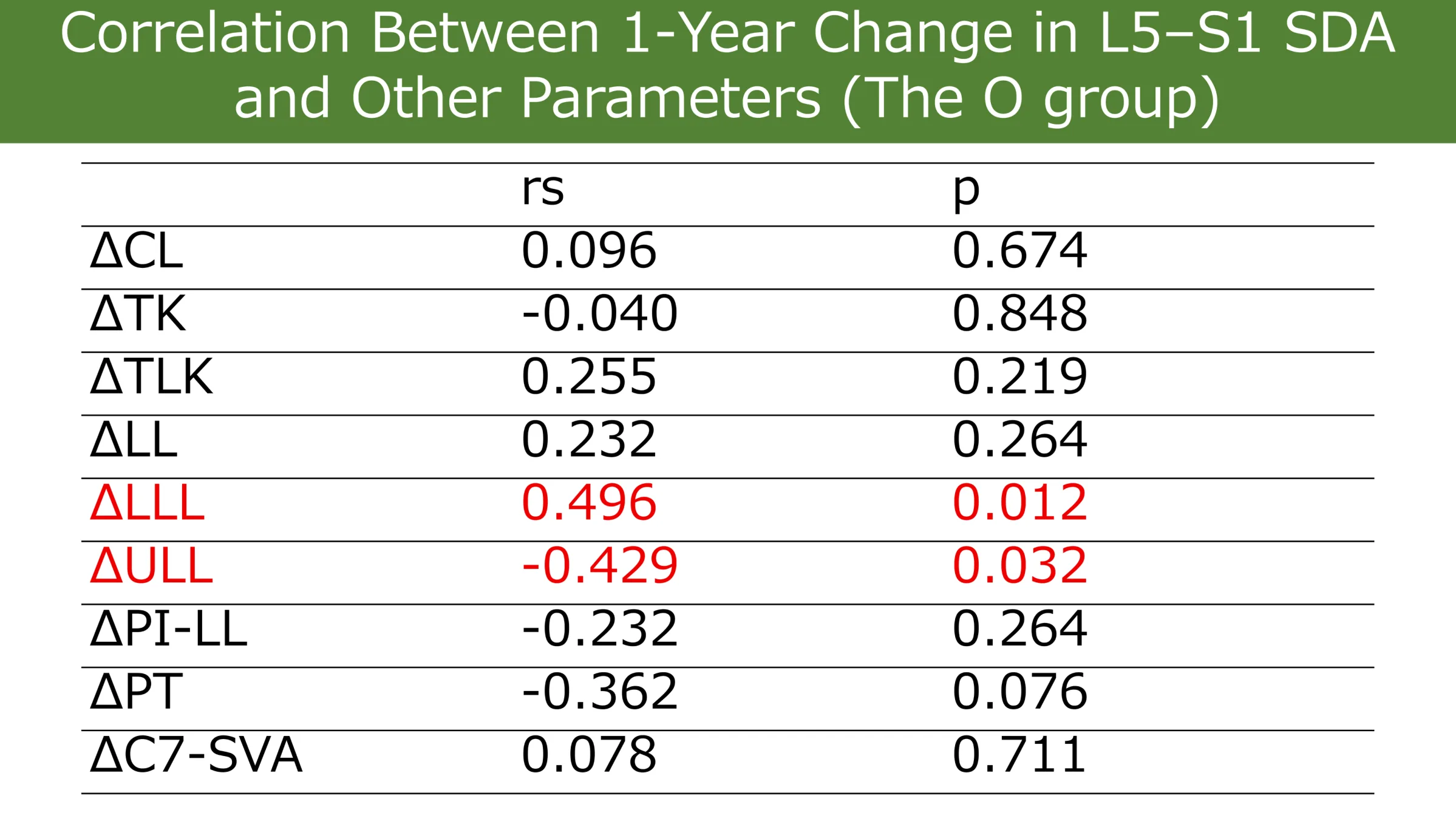
Compared with the T group, the O group showed greater improvements in change in L5-S1 SDA (9.6±5.0° vs 2.9±4.3°, p<0.001), change in LL (6.5±8.3° vs 0.8±6.5°, p=0.038), and change in LLL (9.5±9.1° vs 2.8±7.2°, p=0.008), with a greater decrease in PT (−1.5±4.1° vs 2.2±5.1°, p=0.002). ODI improvement did not differ significantly (23.7±18.5 vs 12.1±18.2, p=0.188). Compared with the C group (change in L5-S1 SDA=5.9±2.7), the L group (=14.3±2.8) demonstrated a larger change in LLL (14.9±10.2° vs 5.3±5.4°, p=0.014) with a greater reciprocal reduction in ULL (−7.0±6.4° vs 0.1±5.9°, p=0.013). ODI improvement was not significantly different (18.3±14.9 vs 27.0±20.4, p=0.515). In the O group, change in L5-S1 SDA correlated positively with change in LLL (rs=0.496, p=0.012) and negatively with change in ULL (rs=−0.429, p=0.032).
The O group achieved superior lumbosacral segmental lordosis restoration compared with the T group and favorably influenced spinopelvic parameters. Greater L5–S1 correction increased LLL and induced reciprocal reduction of ULL. Although short-term clinical outcomes were similar, suppressing upper lumbar hyperlordosis may reduce stress on adjacent segments and provide potential long-term benefits.
Figures and tables
As submitted with the abstract. Tap a figure to open it at full size.