
Cervical & Neural Pathologies
Preserving Image Quality with Ultrafast Cervical Spine MRI Using HASTE and Deep Learning–Based Reconstruction
- Nakamura Memorial Hospital, Sapporo, Japan
Abstract
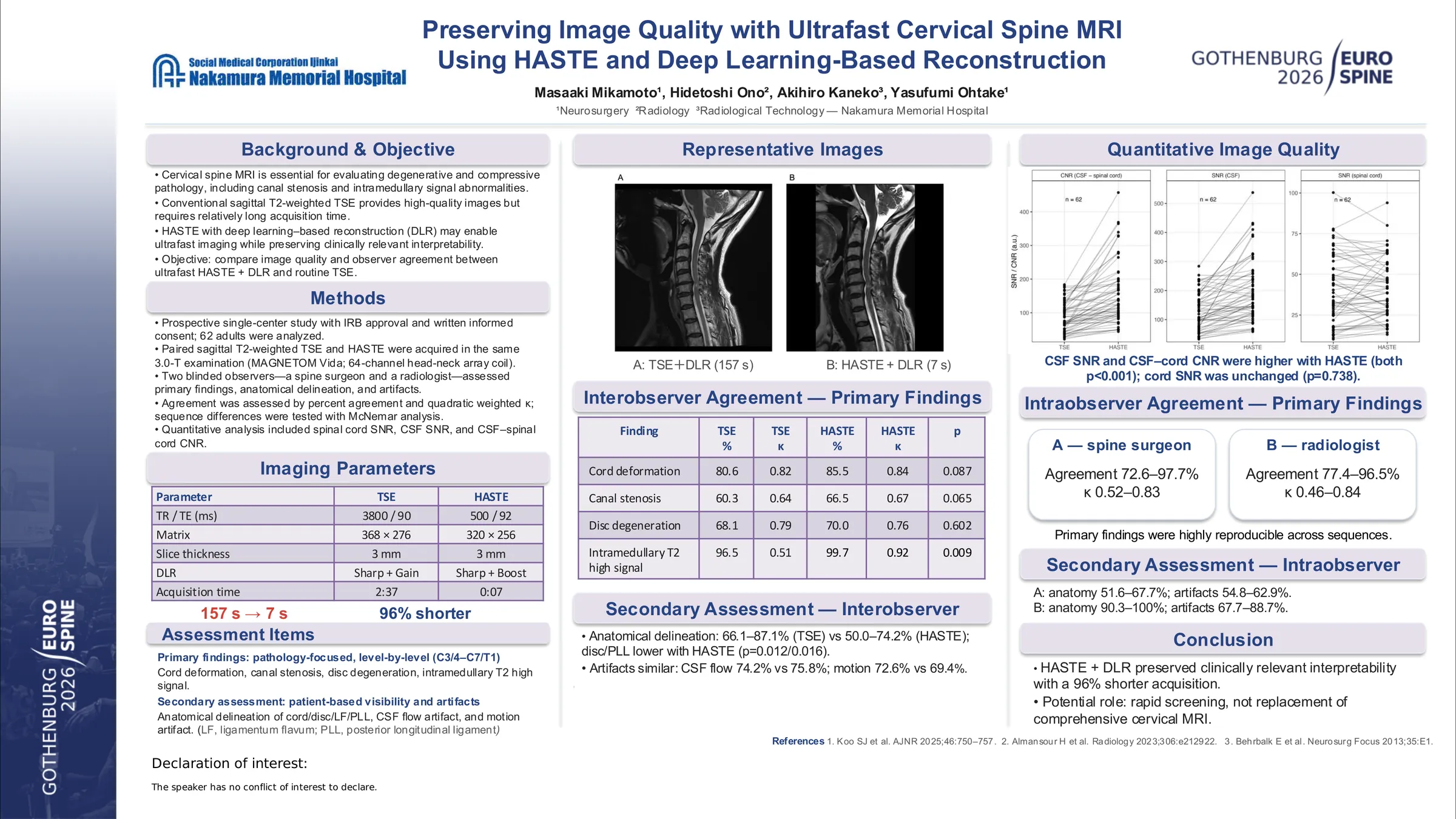
Ultrafast MRI techniques may reduce examination time and improve workflow in cervical spine imaging; however, potential degradation of diagnostic image quality remains a concern. Half-Fourier acquisition single-shot turbo spin echo (HASTE) combined with deep learning–based reconstruction may enable rapid cervical spine MRI while preserving clinically relevant image quality. This study compared image quality and observer agreement between ultrafast HASTE with deep learning–based reconstruction and conventional turbo spin echo (TSE).
Sixty-two adult patients underwent sagittal T2-weighted cervical spine MRI using both conventional TSE and HASTE within a single examination. HASTE was acquired with deep learning–based reconstruction with an acquisition time of approximately 7 seconds. Two independent observers performed visual assessments of primary imaging findings, anatomical delineation, and image artifacts, and interobserver and intraobserver agreement were evaluated. Quantitative image quality was assessed using signal-to-noise ratio (SNR) of the spinal cord and cerebrospinal fluid (CSF), and contrast-to-noise ratio (CNR) between CSF and the spinal cord.
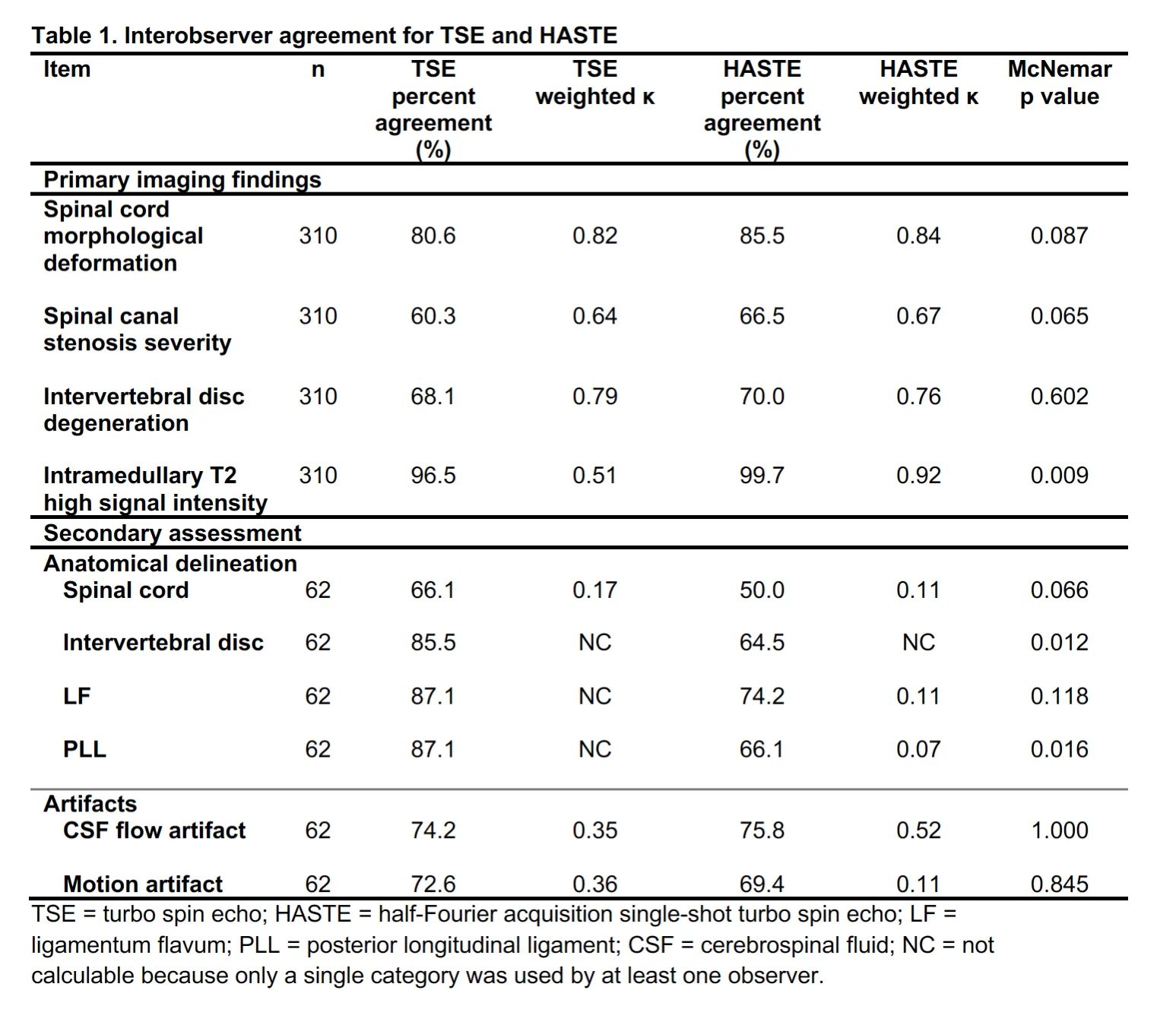
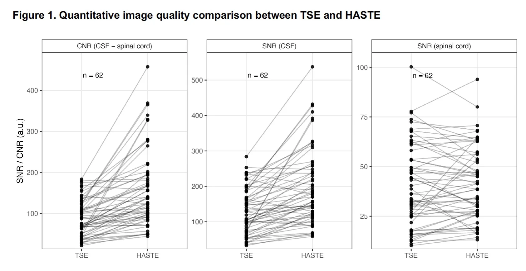
For primary imaging findings, including intervertebral disc degeneration, spinal canal stenosis, and intramedullary T2 hyperintensity, interobserver agreement was generally moderate to high for both sequences, with weighted kappa values in the moderate-to-excellent range. Agreement for anatomical delineation showed structure- and observer-dependent variability, with generally lower kappa values than those for primary imaging findings. Assessment of CSF flow and motion artifacts showed similar trends between sequences. Importantly, images rated as non-diagnostic were rare for both sequences, indicating preserved diagnostic acceptability even with ultrafast acquisition. Quantitative analysis demonstrated significantly higher CSF SNR (196.3 ± 105.7 vs 128.3 ± 64.1) and higher CSF–spinal cord CNR (155.5 ± 91.3 vs 88.0 ± 44.3) for HASTE than for TSE (both p < 0.001), while spinal cord SNR did not differ significantly between sequences (p = 0.738).
Despite a markedly shorter acquisition time, HASTE with deep learning–based reconstruction preserved diagnostic acceptability, quantitative image quality, and observer agreement comparable to those of conventional TSE, supporting its potential utility as an ultrafast sequence for cervical spine MRI screening.
Figures and tables
As submitted with the abstract. Tap a figure to open it at full size.