
Adult Deformity
Pre-postoperative reciprocal changes for ASD patients with long thoracolumbar fusion
- Arts et Métiers ParisTech, Paris, France
- Rouen University Hospital, Rouen, France
- Saint Joseph University of Beirut, Beirut, Lebanon
- Rouen Normandy University, Rouen, France
- Hôpital P. Wertheimer, Hospices Civils de Lyon, Lyon, France
- CHU Bordeaux Pellegrin, Périgueux, France
- Hôpital Européen Georges Pompidou, Paris, France
- Hôpital Privé du Dos Francheville, Périgueux, France
- Saint Joseph University of Beirut, Bayrut, Lebanon
- Research fund onclinical biomecanics of the spine, Paris, France
Abstract
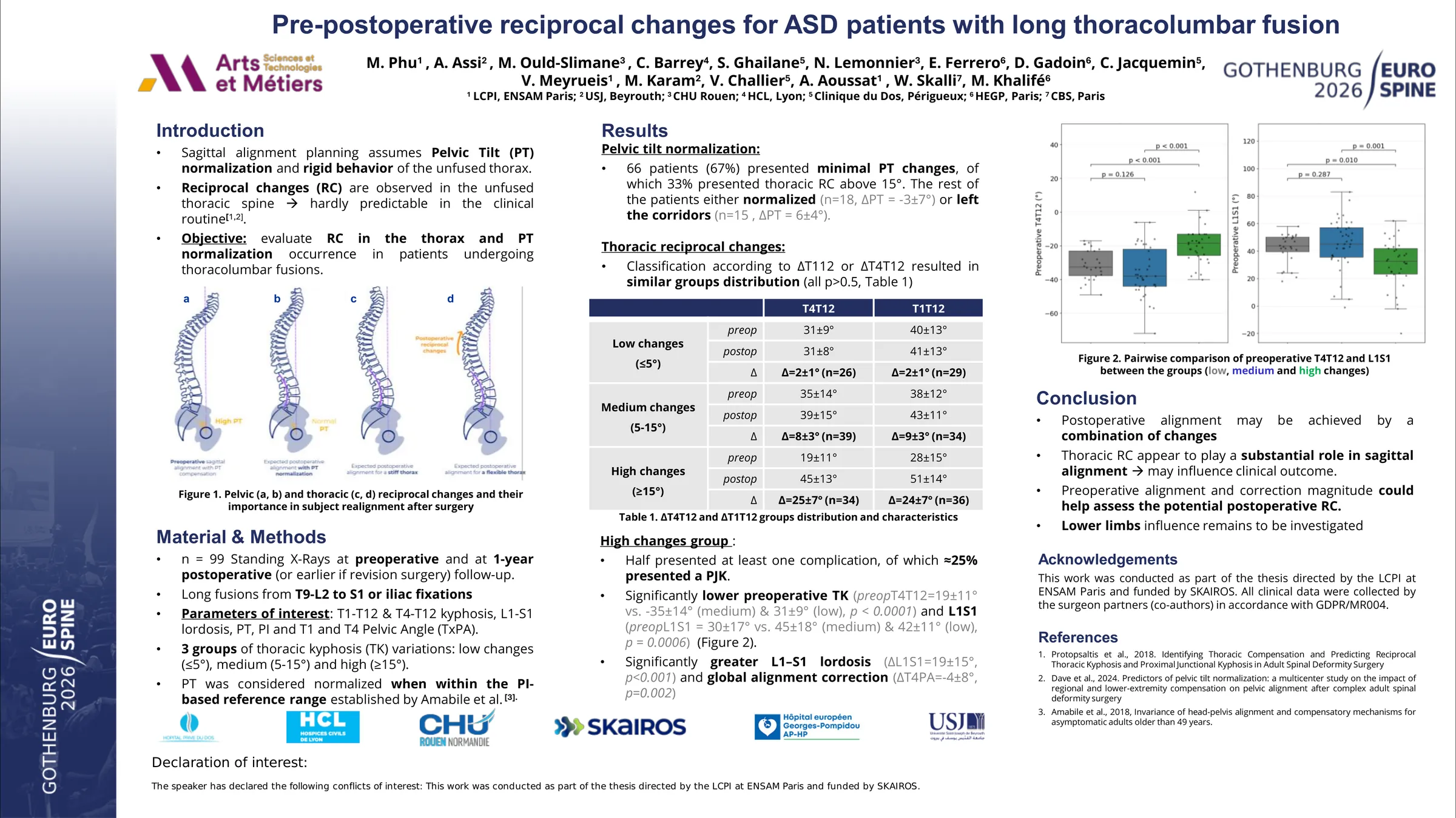
Several surgical planning algorithms aim to achieve postoperative sagittal alignment by computing the target curvature of the instrumented segment, with the assumption of Pelvic Tilt (PT) normalization in relation with the Pelvic Incidence (PI) and that the unfused thorax acts as a rigid ensemble. However, these models often assume a predictable postoperative pelvic compensation and neglect reciprocal changes occurring in the non-fused thoracic spine. Indeed, there are still only few studies on the real postoperative behavior of the unfused spine. The aim of this study was to evaluate postoperative reciprocal changes and pelvic tilt normalization occurrence in patients undergoing long fusions.
In this multicentric retrospective study, patients included were those undergoing fusions extending from the thoracolumbar junction (T9 or below) to sacral or sacroiliac fixations. Preoperative and 1-year postoperative sagittal X-Rays (or earlier if revision surgery was necessary) were analyzed. Parameters of interest included the T4-T12 kyphosis, L1-S1 lordosis (LL), PT, PI and the PI-LL mismatch. T4-T12 pre and post-operative variations (∆T4T12) were classified into three groups: low changes (∆T4T12 < ± 5°), medium changes (between 5 and 15°) and high (∆T4T12 ≥ ±15°). Pelvic tilt was considered normalized when within the theoretical PI-based reference range.
A total of 99 patients were included (69 females, mean age 66.1 ± 10.2 years old, mean BMI 26.7 ± 5.0 km/m²). For the low ∆T4T12 group (n=23), the mean change was 2.13 ± 1.28° versus 8.32 ± 2.76° for medium changes (n=35). 42 patients presented a high ∆T4T12 variation, of which 28% (n=12) had a PJK. Patients in the high ∆T4T12 group had a significantly greater L1–S1 lordosis correction than both other groups (p < 0.005). ΔPI–LL was significantly higher for the high ∆T4T12 group (p ≈ 0.00001). 47% of patients (n=47) presented minimal PT changes (∆PT<±5°) and only 6% presented changes above 15°. Regarding the PI-specific reference corridors, 70 patients did not normalized or worsened, of which 42% presented high ∆T4T12 changes and 28% medium ones.
These findings suggest that postoperative alignment is not systematically achieved through pelvic compensation alone. Reciprocal thoracic changes appear to play a substantial role in sagittal alignment and may influence mechanical outcome. Further research is needed to develop reliable clinical assessment of the thoracic spine flexibility and pelvic compensatory potential prior to the surgery, which may improve the accuracy and individualization of surgical alignment planning.