
Adult Deformity
Long fusion with pelvic fixation in ASD lead to frontal instability during gait
- Saint Joseph University of Beirut, Beirut, Lebanon
- Research fund on clinical biomechanics of the spine, Paris, France
Abstract
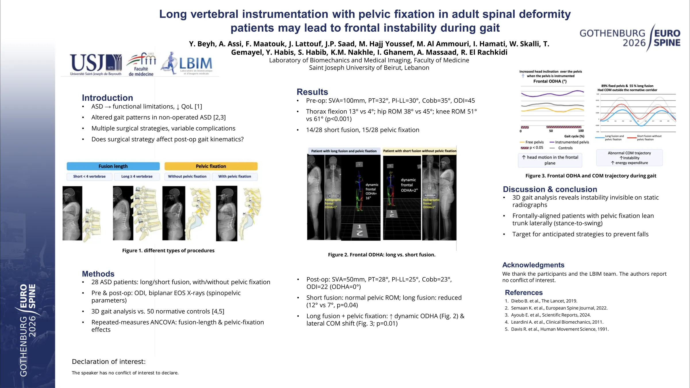
ASD patients are known to present with functional limitations related to their deteriorated QOL. In the recent years, 3D movement analysis allowed for the detailed description of strategies of walking and gait stability in non-operated ASD, depending on the severity of their spinal deformity. Surgery is proposed in some cases with high rate of complication and/or persistent postoperative pain and discomfort. This introductory study aims to evaluate functional stability during gait in post-operative ASD patients, while considering the type of surgery.
ASD patients scheduled for surgery participated in this study. All patients underwent surgery with either long (≥4 levels) or short (<4) fusion, with or without pelvic fixation. Pre and postoperative examination included: ODI questionnaire, biplanar X-rays with calculation of spinopelvic parameters, and 3D gait analysis with calculation of trunk, pelvis and lower limb kinematics. Variables were compared using a mixed model while evaluating the effect of fusion length and pelvic fixation.
28 ASD patients were included. Preoperatively, patients had sagittal malalignment (SVA 100mm, PT: 32°, PI-LL: 30°) and/or frontal spinal deformity (Cobb: 35°). They had an average ODI of 45. Among the 28 patients, 14 had short fusion while 15 had pelvic fixation. The mean follow-up period was 1.6 years (6 to 30 months). Postoperatively, radiographic parameters were as follow in patients with sagittal malalignment SVA: 50mm, PT: 33° and PI-LL: 25°, and Cobb: 20° for patients with frontal scoliosis. All patients were frontally aligned (frontal ODHA= 0°). ODI score was reduced to 22.
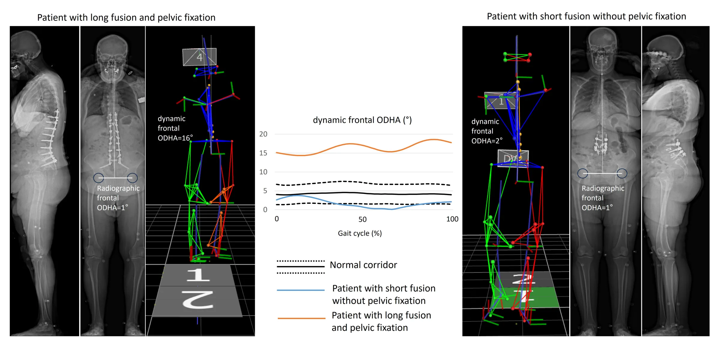
On the dynamic level, all patients demonstrated comparable dynamic sagittal alignment. The short fusion group maintained a normative pelvic range of motion in the horizontal plane, compared to the long fusion group (12 vs 7°, p=0.04). Patients having a long fusion, and especially those with pelvic fixation, showed an increased frontal instability while walking (dynamic frontal ODHA: 6.5 vs 3°, p=0.04; Figure 1). In fact, 89% of patients with fixed pelvis and 55% of those with long fusion had their center of mass (COM) shifted laterally outside the normative corridor (Chi-squared test: p=0.01).
Even on a small sample of patients followed up after surgery, 3D functional evaluation seems to detect differences in patient’s strategies during walking. This introductory study revealed for the first time that patients with pelvic fixation, and to a lesser extent with long fusion, who were frontally aligned on the radiographs, need to lean their trunk laterally, outside of the normative zone, when transferring their weight to the supporting limb during the stance to swing walking transition. This behavior may reflect dynamic instability during walking, where rehabilitation could be crucial in teaching anticipated strategies to prevent falls.
Figures and tables
As submitted with the abstract. Tap a figure to open it at full size.